Showing the invisible: New research to help us see bacteria in the body
Feature Image

In recent years, research has increasingly shown us the importance of bacteria and other microbes in the human microbiome for maintaining health. Now, researchers at Lawson Health Research Institute are pioneering new imaging methods to see these microbes in the human body and open new avenues for health research. Early results of preclinical studies at Lawson have found positron emission tomography-magnetic resonance (PET/MR) imaging could allow the tracking and identification of bacteria inside the body and lead to more targeted use of antibiotic treatments.
Accurate targeting of antibiotic treatments can prevent antimicrobial resistance – when bacteria, viruses, fungi and parasites no longer respond to medication. According to a United Nations report, it is estimated that by 2050, antimicrobial resistance could result in 10 million deaths each year – more than cancer. New imaging research could be a gamechanger for treating bacterial infections by allowing us to see bacteria in the body using medical imaging equipment and then targeting the bacteria with specific therapies.
“Traditional imaging of infection means that you're looking at tissue damage; the bacteria have already started the process of inflammation and are wreaking havoc,” explains Dr. Donna Goldhawk, Lawson Scientist at St. Joseph’s Health Care London (St. Joseph’s). “Imaging bacteria catches the infection at an earlier stage. When you can image a particular species of bacteria, you can narrow the type of antibiotic that you might want to treat it with – reducing the need for broad-spectrum antibiotics that can lead to antimicrobial resistance.”
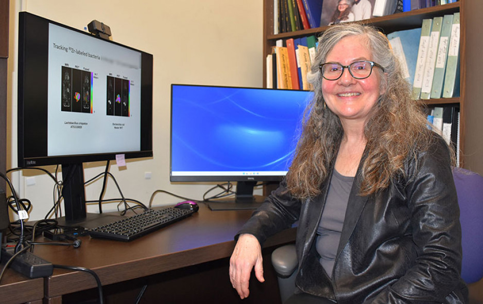
Imaging bacteria using PET/MR technology begins with attaching tracer molecules – also called isotopes – to specific bacteria in order to follow the movement of the microbes. A recent study from Lawson used PET/MR imaging to track bacteria labelled with an isotope called Zirconium-89 (89Zr) in a preclinical model. The researchers were able to demonstrate that PET/MR imaging could track ingested bacteria through the gut.
“Imaging of bacteria is a very new application of how PET/MR technology can be used. Using isotopes like 89Zr to label bacteria would allow you to image the same individual repeatedly and follow the ingestion of specific bacteria from the stomach through the digestive system since those isotopes last a long time,” adds Dr. Frank Prato, Scientist at Lawson and Lead of the Lawson Imaging research program.
This also has the potential to allow imaging of bacteria that migrate to other areas of the body like the brain, bladder, kidneys, and reproductive system. In the future, this technology could allow researchers to identify specific bacteria present and target those bacteria.
Similar research is underway to examine whether bacteria without tracers can also be tracked using MR imaging based on differences in their characteristics, like associations with specific metals. This could allow imaging of specific bacteria in the gut and how they respond when gut infections are treated with antibiotics, probiotics or microbial therapies like fecal microbiota transplantation (FMT).
With or without tracer molecules, both imaging methods could eventually become important for improving the efficacy and wider implementation of FMT, which introduces healthy microbes from donors into a patient’s gut with the goal of having the healthy bacteria reinstate a balanced microbiome. FMT is currently used to treat recurrent infections of C. diff. (Clostridium difficile), but new applications are expanding with clinical trials looking at its use to treat a variety of diseases, including certain forms of cancer. The ability to see how the balance of bacteria is changing could accelerate the development of effective new therapies.
While more research is needed, these studies are moving the monitoring of bacteria using PET/MR imaging closer to clinical implementation. The research has been made possible in part thanks to collaborations with Siemens Healthineers, Cubresa Inc. and London X-Ray Associates.