Search
Search
1347 Search Results:
… swelling, redness, stiffness, and pain. Depending on the areas affected and functional ability, either a sedan or van … swelling, redness, stiffness, and pain. Depending on the areas affected and functional ability, either a sedan or van …
Parkwood Hospital Driving Assessment and Rehabilitation Program 801 Commissioners Road East London, Ontario N6C 5J1 Phone: (519) 685-4070 Fax: (519) 685-4576 DRIVING WITH RHEUMATOID ARTHRITIS Rheumatic disease includes nearly 100 different conditions, which cause pain in the joints and connective ti...
Rheumatology Infusion Room LOCATION AND HOURS The Rheumatology Infusion Room is located within the Rheumatology Clinic at St. Josephs Hospital: in Zone D, on Level 2. You will check in at the same reception area as when you see your rheumatologist. Hours of Operation: Monday to Friday (excluding sta...
RHINOPLASTY INTRODUCTION The term rhinoplasty refers to surgery performed to alter the structure of the external nose. It is commonly referred to as a nose job. When combined with surgery on the nasal septum it is referred to as a septorhinoplasty. This may be performed for purely cosmetic reasons ...
June 16, 2020; Page 1 of 2 Sacroiliac Joint Injection What is a sacroiliac (SI) joint injection and why is it performed? The SI joints connect the spine to the pelvis and allow for motion. There are two SI joints: one on the left and one on the right. The SI joint is surrounded by a lubricant fille...
Scientists studying carbon monoxide as a possible treatment for sepsis
Sepsis is the leading cause of death worldwide with limited treatment options
MEDIA RELEASE
For immediate release
LONDON, ON – Sepsis is a life-threatening condition that occurs when the body’s response to an infection triggers excessive inflammation. The inflammatory response can cause damage to organs such as the heart, liver, lungs and brain. While there are currently limited treatments for sepsis, researchers at Lawson Health Research Institute are working to change that by examining the use of carbon monoxide-releasing molecules to treat patients. “This is an unusual approach that is looking at using carbon monoxide which is the infamous gas molecule,” says Dr. Gediminas Cepinskas, Scientist and Director of the Centre for Critical Illness Research at Lawson. “If administered and used in small non-toxic concentrations, carbon monoxide can offer very potent protective and anti-inflammatory effects.”
In studies on the subject, including the most recent one published in the journal of Experimental Biology and Medicine, the research team was able to demonstrate efficacy in using carbon monoxide-releasing molecules to protect individual cells in the liver and lungs of sepsis induced inflammation in preclinical models. “We have been working on isolated organs and organ specific cells to test carbon monoxide-releasing molecules to narrow down which specific cells are more sensitive to treatment and which biochemical pathways are involved,” Says Dr. Cepinskas. “We are making great progress in our work and have had success in addressing the efficacy of carbon monoxide-releasing molecules in preclinical models.”
Dr. Cepinskas is one of just a few scientists worldwide studying carbon monoxide-releasing molecules to treat inflammatory conditions such as sepsis. While carbon monoxide is commonly known as dangerous, using it in a controlled manner does not pose a danger and may have therapeutic potential. “Our immune system is usually our defense system, but with sepsis it becomes so activated it starts to attack our own cell tissues, resulting in injury and dysfunction of the affected organs,” explains Dr. Cepinskas. “Almost each and every cell in our body naturally produces carbon monoxide which is used in defense against harmful and injured stimuli. We are taking advantage of this knowledge and currently we are the only lab in Canada working on this potential carbon monoxide-based therapy.”
Dr. Cepinskas is also collaborating with clinicians at London Health Sciences Centre (LHSC) to study the use of carbon monoxide-releasing molecules to treat limb compartment syndrome and to improve organ transplantation.
The research team, which has patents in the area of carbon monoxide-releasing molecules, is now working with the pharmaceutical industry to move this potential therapy into human clinical trials in the future.
-30-
About Lawson Health Research Institute
Lawson Health Research Institute is one of Canada’s top hospital-based research institutes, tackling the most pressing challenges in health care. As the research institute of London Health Sciences Centre and St. Joseph’s Health Care London, our innovation happens where care is delivered. Lawson research teams are at the leading-edge of science with the goal of improving health and the delivery of care for patients. Working in partnership with Western University, our researchers are encouraged to pursue their curiosity, collaborate often and share their discoveries widely. Research conducted through Lawson makes a difference in the lives of patients, families and communities around the world. To learn more, visit www.lawsonresearch.ca.
Media Contacts
Celine Zadorsky
Senior Media Relations Consultant
Communications & Public Engagement
T: 519-685-8500 ext. 73502
Celine.zadorsky@lhsc.on.ca
… Schizophrenia is a psychiatric disorder that can affect all areas of daily life including one’s ability to drive. The … attention, reaction time, and sensorimotor performance areas. For example, sedation is a common side effect from … aware of one’s strengths and limited capabilities in these areas when considering operating a vehicle. Common factors …
Parkwood Hospital Driving Assessment and Rehabilitation Program 801 Commissioners Road East London, Ontario N6C 5J1 Phone: (519) 685-4070 Fax: (519) 685-4576 DRIVING WITH SCIZOPHRENIA Schizophrenia is a psychiatric disorder that can affect all areas of daily life including ones ability to drive. The...
… to the appropriate departments for follow up? Patient Care Areas: 1) Have issues regarding barriers been identified … 'this' user population incorporated into the design of new areas? Wayfinding / Volunteers: Annual Accessibility Plan … to the appropriate departments for follow up? Patient Care Areas: 1) Have issues regarding barriers been identified …
Annual Accessibility Plan regarding ONTARIANS WITH DISABILITIES ACT for the St. Josephs Health Care, London September 2003 - August 2004 Submitted to Cliff Nordal Chief Executive Officer 30 September 2003 Prepared by Nick Kokkoros Facilitator Accessibility Working Group This publication is available...
… Draft: July, 2004 11 o) Interpreters Brief Description: All areas of SJHC are advised to provide trained interpreters … Number of accessible entrances close to parking areas and clinics. Architectural Some washroom stalls are … access some diagnostic / ambulatory outpatient and clinical areas. Annual Accessibility Plan Draft: July, 2004 15 9. …
Annual Accessibility Plan for the St. Joseph Health Care, London September 2004 - August 2005 Submitted to Cliff Nordal Chief Executive Officer 30 September 2004 Prepared by SJHC Accessibility Working Group Co-ordinator, Derek Lall This publication is available on the hospitals website and in altern...
… Rarely, some patients can have persistent numbness of these areas. Septal perforation: A septal perforation is a hole in … Rarely, some patients can have persistent numbness of these areas. Septal perforation: A septal perforation is a hole in …
SEPTOPLASTY & TURBINATE SURGERY INTRODUCTION Many people have difficulty with nasal congestion and stuffiness. When someone has chronic stuffiness, theyre often forced to breathe through their mouth, leading to a sensation of a dry mouth. In many patients, these symptoms get worse at night when they...
… to do so, complete patrols of the grounds and public access areas to identify possible hazards 4.1.3. Report potential … to do so, complete patrols of the grounds and public access areas to identify possible hazards 4.1.3. Report potential …
~ SIJOSEPHs HEALTH CARE LONDON CORPORATE Procedure: SEVERE WEATHER Owner of Procedure: Emergency Management and Risk Specialist Approval by: Emergency Management Committee Date: 2022-06-02 Original Effective Date: 2021-05-03 Reviewed Date(s): 2022-05-27 Revised Date(s): 2022-05-27 PURPOSE Adverse or...
Parkwood Institute Main Building Sexual Health after a Brain Injury This pamphlet contains information for patients living with a brain injury, including: How sexual health may be affected after a brain injury How to cope with changes to sexual health following a brain injury Tips for being sexua...
Parkwood Institute Main Building Sexual Health After Spinal Cord Injury 1 Parkwood Institute Main Building Sexual Health After Spinal Cord Injury 2 Spinal Cord Injury Rehabilitation Program You can do it, we are here to help! Sex and sexuality are important parts of being human, however after a spin...
Parkwood Institute Main Building Sexual Health After Stroke Sexual Health After Stroke Stroke Rehabilitation Program Page 2 You can do it, we are here to help! Sex and sexuality are important parts of being human. After having a stroke, many things can change. The way in which you experience your bo...
Parkwood Institute Main Building Sexual Health After Stroke Sexual Health After Stroke Stroke Rehabilitation Program Page 2 You can do it, we are here to help! Sex and sexuality are important parts of being human. After having a stroke, many things can change. The way in which you experience your bo...
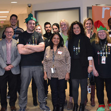
Sharing the gift of hope through research
Community members and those in the field of health research attended a special open house and interactive tour by research groups at Lawson Health Research Institute – celebrating the holiday spirit and the gift of hope that is made possible through hospital-based research.
A part of St. Joseph’s Health Care London, Parkwood Institute represents the next era in care, recovery and rehabilitation. Across the site, clinical and research teams in different disciplines and specialties are collaborating in new ways.
The teams that make up Parkwood Institute Research, a Lawson program, are conducting clinical studies with the goal of understanding disease and improving care for a wide range of patients.
“Many people in the community know the high-quality and compassionate care that is supported by the various clinical teams at Parkwood Institute,” says Dr. Cheryl Forchuk, Beryl and Richard Ivey Research Chair in Aging, Mental Health, Rehabilitation and Recovery, and Assistant Scientific Director at Lawson. “What most don’t know is that we have research teams working across these sites, with each other and research patients.
Researchers tackle the most important challenges and provide access to highly innovative and meaningful solutions that improve the lives of patients and their families, added Dr. Forchuk.
Image
At the open house on November 30, there were 11 interactive displays in the areas of cognitive vitality and brain health, mobility and activity and mental health. This included the Gait and Brain Laboratory, the Operational Stress Injury Clinic, wound care, the Mental Health Nursing Research Alliance and more:
- The Mental Health INcubator for Disruptive Solutions (MINDS) of London Middlesex is a social innovation lab focused on developing, testing, implementing and evaluating disruptive solutions that promote the mental and emotional wellbeing of Transition-Aged Youth in our London-Middlesex community.
- The Canadian Consortium on Neurodegeneration in Aging (CCNA), supported by CIHR and many partners, is the premier research hub for all aspects of research involving neurodegenerative diseases that affect cognition in aging – including Alzheimer's disease.
- A dynamic lab with the top neurorehabilitation evidence-based reviews in stroke, brain injury, spinal cord injury and multiple sclerosis, or insight into clinical trials of stroke rehabilitation using exoskeletons, exercise paradigms, pharmaceuticals, and clinical studies of psychosocial factors that influence chronic pain in brain injury and spinal cord injury populations.
The open house had a festive theme and each of the exhibits involved a problem solving element to encourage learning and foster teamwork.
Image
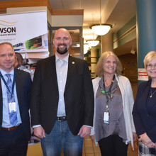
Peggy Sattler, Member of Provincial Parliament for London West, and Terence Kernaghan, Member of Provincial Parliament for London North Centre, were among the over 300 people in attendance.
“This special open house opportunity is meant to be a fun experience where you were also learning about research,” explains Dr. Forchuk. “During this holiday season, we wanted to share our own gift of hope in the form of collaborative research that is making a real difference.”
See photos from the open house on Facebook.
Scientist
~ ROTH I M(;FARLANE ~ ... HAND & UPPER LIMB CENTRE ~~as ST. JOSEPH'S HEALTH CARE LONDON 0Western ~ HealthSciences School of Physical Therapy What You Should Know Before Surgery -Reverse/Total Shoulder Replacement- Sling Use Why is it important? - Helps support your shoulder for comfort and soft t...
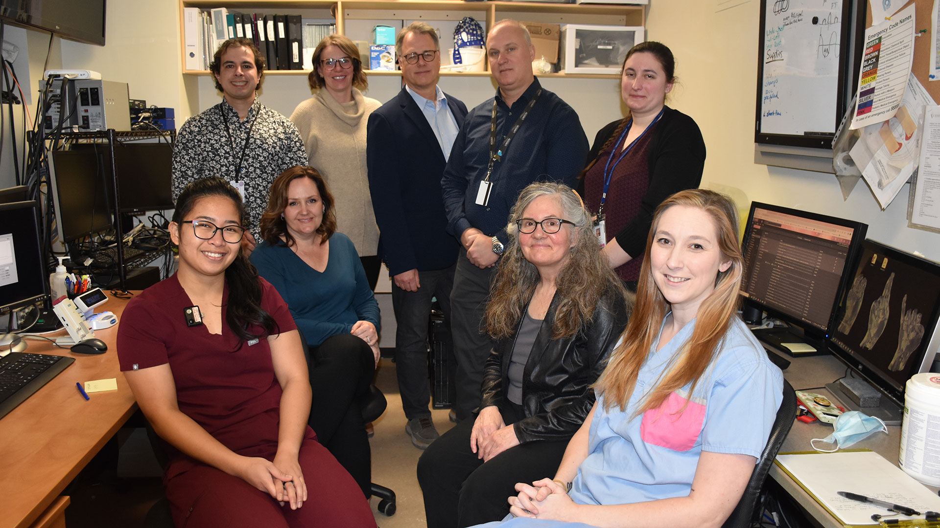
Showing the invisible: New research to help us see bacteria in the body
In recent years, research has increasingly shown us the importance of bacteria and other microbes in the human microbiome for maintaining health. Now, researchers at Lawson Health Research Institute are pioneering new imaging methods to see these microbes in the human body and open new avenues for health research. Early results of preclinical studies at Lawson have found positron emission tomography-magnetic resonance (PET/MR) imaging could allow the tracking and identification of bacteria inside the body and lead to more targeted use of antibiotic treatments.
Accurate targeting of antibiotic treatments can prevent antimicrobial resistance – when bacteria, viruses, fungi and parasites no longer respond to medication. According to a United Nations report, it is estimated that by 2050, antimicrobial resistance could result in 10 million deaths each year – more than cancer. New imaging research could be a gamechanger for treating bacterial infections by allowing us to see bacteria in the body using medical imaging equipment and then targeting the bacteria with specific therapies.
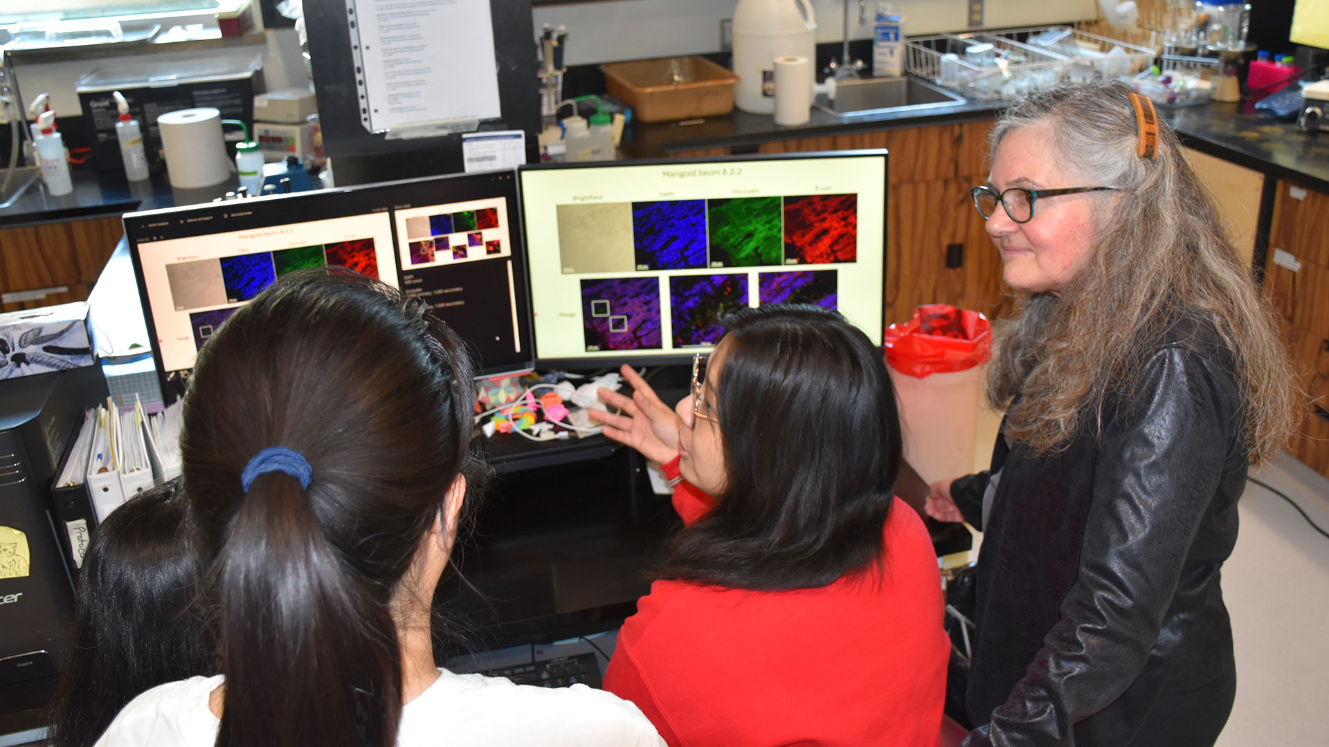
“Traditional imaging of infection means that you're looking at tissue damage; the bacteria have already started the process of inflammation and are wreaking havoc,” explains Dr. Donna Goldhawk, Lawson Scientist at St. Joseph’s Health Care London (St. Joseph’s). “Imaging bacteria catches the infection at an earlier stage. When you can image a particular species of bacteria, you can narrow the type of antibiotic that you might want to treat it with – reducing the need for broad-spectrum antibiotics that can lead to antimicrobial resistance.”
Imaging bacteria using PET/MR technology begins with attaching tracer molecules – also called isotopes – to specific bacteria in order to follow the movement of the microbes. A recent study from Lawson used PET/MR imaging to track bacteria labelled with an isotope called Zirconium-89 (89Zr) in a preclinical model. The researchers were able to demonstrate that PET/MR imaging could track ingested bacteria through the gut.
“Imaging of bacteria is a very new application of how PET/MR technology can be used. Using isotopes like 89Zr to label bacteria would allow you to image the same individual repeatedly and follow the ingestion of specific bacteria from the stomach through the digestive system since those isotopes last a long time,” adds Dr. Frank Prato, Scientist at Lawson and Lead of the Lawson Imaging research program.
This also has the potential to allow imaging of bacteria that migrate to other areas of the body like the brain, bladder, kidneys, and reproductive system. In the future, this technology could allow researchers to identify specific bacteria present and target those bacteria.
Similar research is underway to examine whether bacteria without tracers can also be tracked using MR imaging based on differences in their characteristics, like associations with specific metals. This could allow imaging of specific bacteria in the gut and how they respond when gut infections are treated with antibiotics, probiotics or microbial therapies like fecal microbiota transplantation (FMT).
With or without tracer molecules, both imaging methods could eventually become important for improving the efficacy and wider implementation of FMT, which introduces healthy microbes from donors into a patient’s gut with the goal of having the healthy bacteria reinstate a balanced microbiome. FMT is currently used to treat recurrent infections of C. diff. (Clostridium difficile), but new applications are expanding with clinical trials looking at its use to treat a variety of diseases, including certain forms of cancer. The ability to see how the balance of bacteria is changing could accelerate the development of effective new therapies.
While more research is needed, these studies are moving the monitoring of bacteria using PET/MR imaging closer to clinical implementation. The research has been made possible in part thanks to collaborations with Siemens Healthineers, Cubresa Inc. and London X-Ray Associates.
SINUS TUMORS INTRODUCTION A tumor is a mass or growth. Tumors of the nose and paranasal sinuses are rare. They represent less than 1% of all tumors. These lesions can be either benign (non-cancerous) or malignant (cancerous). They vary in location, size and type. The care for a patient with a sinona...
… The spectacular views, recreational space, and natural areas were thoroughly enjoyed. Since the new site was some … encompass �� sites. Through the newly-defined five role areas, St. Joseph’s now provides a scope of services … to almost every segment of the population. Those role areas include: acute/ambulatory care; specialized mental …
S i S t e r the hiStory of the SiSterS of St. JoSeph of London Special thanks to Lori Perrie and Sister Mary Zimmer of the Sisters of St. Joseph of the Diocese of London for their knowledge, expertise and keen attention to detail. cover: The caring hands of Sister Norita Keenan, who worked at St. J...
… The spectacular views, recreational space, and natural areas were thoroughly enjoyed. Since the new site was some … •33 •3� began with the gathering of lay women in various areas of Southwestern Ontario. These women work together to … The spectacular views, recreational space, and natural areas were thoroughly enjoyed. Since the new site was some …
s i s t e r tHe History of tHe sisters of st. JosepH of london 2 Special thanks to Lori Perrie and Sister Mary Zimmer of the Sisters of St. Joseph of the Diocese of London for their knowledge, expertise and keen attention to detail. cover: The caring hands of Sister Norita Keenan, who worked at St. ...