Search
Search
251 Search Results:
… Director. James Calvin Stephen Wetmore Narinder Paul Body Breast Cardiothoracic Musculoskeletal Neuroradiology … Director. James Calvin Stephen Wetmore Narinder Paul Body Breast Cardiothoracic Musculoskeletal Neuroradiology …
GUIDELINES FOR PHYSICIAN PERFORMANCE MANAGEMENT - Listing of Physician Leaders in the Department/Organization DEPARTMENT DEPARTMENT CHIEFS SITE CHIEFS DIVISION DIVISION CHIEFS/SECTION HEADS/ DIVISION CHAIRS/MEDICAL DIRECTORS OTHER LEADERSHIP ROLE RESIDENCY PROGRAM DIRECTORS COMMENTS Ashraf Fayad (LH...
… It will be a game-changer for patients receiving care in breast, orthopedic and prostate programs at St. Joseph’s … provide nearly 25,000 short stay surgical treatments for breast, urology, lithotripsy (disintegrating kidney stones), … diagnostic tools. Whether it’s an early-stage diagnosis of breast cancer or providing the right treatment for heart …
Back row left: Holly Bridge, Diane Davis-Miller, Barb Simmonds Front row left: Dr. Jennifer Bjazevic, Roger Blum, Leanne Summers, Dr. Hassan Razvi, Surgical Services You make health care innovation possible. Just one of the reasons why YOU HELPED ENHANCE OUR OPERATING ROOMS 20192020 Community Impact...
… to the donor-supported London Tumour Biobank for future breast cancer research 8 RAISED PLANTER BOXES for elderly … REPORT ST. JOSEPH’S HEALTH CARE FOUNDATION 5 ENHANCING BREAST CANCER SCREENING FOR EARLY DIAGNOSIS When patients come to St. Joseph’s Norton and Lucille Wolf Breast Care Centre for suspected breast cancer, their first …
St. Josephs Health Care Foundation 268 Grosvenor Street PO Box 5777 STN B London, ON N6A 4V2 519-646-6085 sjhc.london.on.ca/foundation CHARITABLE REGISTRATION NUMBER: BN 11918 3390 RR0001 facebook.com/stjosephslondon youtube.com/stjosephslondon twitter.com/stjosephslondon Connect with us YOUR SUPPOR...
2023 Media Releases
New study suggests blood plasma proteins hold answers to better understanding long COVID
New study using nuclear medicine and rare isotopes in the fight against cancer
A new study is examining if probiotics can improve outcomes in knee replacement surgeries
Specific type of inflammation may be linked to risk of colorectal cancer
Study aiming to slow cognitive decline in older adults gets $1.5M
Lawson study validates new biopsy method for breast cancer patients|
New study shows technology could play an important role in mental health support
Precise, high-dose radiation safe and effective for inoperable kidney cancer, study suggests
Could online programming reduce barriers for those with mobility impairments?
London researchers working on a proactive approach to inclusiveness in the classroom
New robotic 3D ultrasound may improve accuracy of liver cancer ablation therapy
… Light Miracle Whip Mediterranean Glazed Haddock Chicken Breast Mediterranean Glazed Haddock GF Sliced Beef Sandwich … of Potato Soup Entrées GF Hawaiian Chicken & Rice Chicken Breast GF Beef Sandwich with Mustard Mediterranean Glazed Haddock GF Chicken Salad Sandwich Chicken Breast Cheese Omelette Salmon Salad in a Dish GF Peanut …
Mount Hope Core Menu REGULAR TEXTURE MENU Effective June 13, 2022 WEEK 1 Monday - Day 1 Tuesday - Day 2 Wednesday - Day 3 Thursday - Day 4 Friday - Day 5 Saturday - Day 6 Sunday - Day 7 BREAKFAST Beverages Apple Juice, Orange Juice, 2% Milk, Coffee, or Tea Cold Cereal Rice Chex Rice Chex Rice Chex R...
3D imaging technology could improve outcomes for patients with breast cancer
A study at Lawson is looking to determine if digital breast tomosynthesis, a type of 3D imaging, is better at detecting breast tissue abnormalities than the 2D mammography regularly used today.
During a conventional digital 2D mammogram, two x-ray images are taken of the breast, one from top-to-bottom and another from side-to-side at an angle. This technology is limited by the overlapping breast tissue that occurs from the required compression of the breast, and breast abnormalities may be hidden.
Image
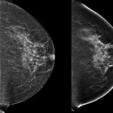
(Left) 2D mammogram image of left breast, where no lesion was visible. (Right) 3D tomographic image of the same breast showing a lesion, indicated by arrows.
A tomosynthesis exam is relatively new technology in which the x-ray tube moves in an arc over the compressed breast and captures multiple images from different angles. The images are then reconstructed into a set of 3D images by a computer. By being able to examine the breast at multiple layers of depth, the radiologist is better able to distinguish normal breast tissue from potential abnormalities. It is therefore assumed that tomosynthesis may solve some challenges associated with standard mammography, and could be especially useful for women with dense breast tissue.
In the Tomosynthesis Mammographic Imaging Screening Trial (TMIST), women are randomized to receive screening with standard digital 2D mammography, or digital 2D mammography plus tomosynthesis. Participants will undergo either an annual or biennial screening frequency, depending on their risk factors for breast cancer, for approximately four years. Then participants will undergo long-term follow-up for at least three more years. Researchers hope this study will help radiologists evaluate whether the newer technology of tomosynthesis is indeed a more effective tool for detecting aggressive tumours.
Through the Ontario Breast Screening Program (OBSP), women between the ages of 50 and 75 receive regular notices through the mail, encouraging them to schedule a mammogram for breast cancer screening. Women scheduled for a regular OBSP breast exam at St. Joseph’s Hospital London (St. Joseph’s) receive a letter with the study’s contact information. Eligible participants are enrolled at the time of their scheduled appointment. Participating in the study does not significantly change the overall experience of the breast exam.
“Our goal is to contribute to the body of evidence around tomosynthesis technology, and ultimately, we hope to improve the outcomes for women diagnosed with breast cancer, meaning, earlier detection,” says Dr. Anat Kornecki, Lawson Scientist and Radiologist at St. Joseph’s.
The TMIST study is being conducted in over 100 centres across Canada, the United States, and Argentina. Approximately 165,000 participants will be recruited.
Scientist
… More women die from osteoporotic fractures than from breast and ovarian cancer combined. Acute and long-term care … of osteoporosis or fractures • chemotherapy (especially for breast cancer), resulting in ovarian failure • chronic use … More women die from osteoporotic fractures than from breast and ovarian cancer combined. Acute and long-term care …
Facts about Osteoporosis What is osteoporosis? Osteoporosis means porous or brittle bones. Weak bones can break easily; fractures of the hip, wrist and spine are commonly associated with osteoporosis. Bone is surprisingly dynamic. It is constantly remodelled; bits of bone are eaten away or resorbed,...
EVERYTHING YOU WANTED TO KNOW ABOUT DIABETES MEDICATIONS MAY 4, 2018 David Leeson R.Ph., B.Sc., B.Sc.Pharm., CDE St. Josephs Health Care London http://www.google.com/url?sa=i&rct=j&q=&esrc=s&source=images&cd=&cad=rja&uact=8&ved=2ahUKEwiQ3vHf8MjaAhUT0IMKHRP-BWUQjRx6BAgAEAU&url=http://starwars.wikia.c...
An image of the future: Innovations in imaging research
Lawson Health Research Institute (Lawson) has long been a leader in biomedical imaging. The first Canadian magnetic resonance imaging (MRI) of a human occurred at St. Joseph’s Health Care London (St. Joseph’s). The country’s first positron emission tomography/computed tomography (PET/CT) and positron emission tomography/magnetic resonance imaging (PET/MRI) scanners were also installed at St. Joseph’s. New developments in imaging research continue to enhance the diagnosis, prevention and treatment of a wide range of diseases, from cancer to post-traumatic stress disorder.
On May 23, Lawson hosted a Café Scientifique event where a panel of Lawson Imaging scientists discussed their cutting-edge work. Guests had the opportunity to ask questions as part of an open-forum discussion to gain insights from the speakers, and from one another.
In celebration of Canada’s 150th anniversary as a nation, this event is the first of a two-part series focusing on the future vision for health care in Canada and the legacy that research at Lawson will leave.
Imaging of the heart: Seeing the cause of chest pain more clearly
By Dr. Ting-Yim Lee, Lawson scientist, Medical Physicist at St. Joseph’s, professor at Western University’s Schulich School of Medicine & Dentistry, and scientist at Robarts Research Institute
When patients with chest pain arrive in the emergency department, they are given an electrocardiogram (ECG) and blood test. These diagnostic tests determine if the pain has a non-cardiac cause (such as heart burn), if it is caused by a heart attack, or if the patient has angina (plaque formation in the coronary arteries that either reduces or temporarily cuts off blood flow to the heart) but did not have a heart attack.
If a patient has angina, they are then given additional diagnostic testing to see whether a blood clot has formed and where it is located. This is determined by two different imaging techniques: x-ray imaging (angiogram) and nuclear imaging. This process is invasive and means that patients must be scheduled for two different exam days. Using two techniques also means that there can be image misalignment, and the images often provide poor detail.
Dr. Ting-Yim Lee’s lab has pioneered a Computed Tomography (CT) method for imaging blood flow to the heart muscle (CT Perfusion), which can help patients avoid unnecessary tests and treatment, as well as reduce health care costs.
“CT imaging is a non-invasive imaging technique that uses x-rays to create high-detail cross-sectional images of the body. Using this method, we can evaluate the degree of blockage in coronary arteries – with one diagnostic test instead of two,” says Dr. Lee.
Using light and sound to improve breast surgery
By Dr. Jeffrey Carson, Lawson scientist and associate professor at Western University’s Schulich School of Medicine & Dentistry
“Most women diagnosed with breast cancer undergo surgery, and months of chemotherapy and radiotherapy. They must deal with the discomfort, side-effects, emotional stress and financial burden of treatment. Almost one in four surgeries for breast cancer must be repeated, meaning many women have to go through this all over again,” says Dr. Jeffrey Carson.
In breast conserving surgery, there is a high chance of repeat surgery as the surgeon must see and remove 100 per cent of the tumour in order for it to be successful. They are not able to determine whether the entire tumour was removed until after the surgery has been completed.
Dr. Carson and his team at St. Joseph’s have developed a technology called Intraoperative Photoacoustic Tomography (iPAT), which has the potential to reduce the chance of repeat surgery for breast cancer. The technology is able to image surgery specimens in the operating room during surgery, allowing surgeons to determine whether the whole tumour has been removed before the surgery is complete.
How imaging can improve the management of epilepsy
By Dr. Udunna Anazodo, postdoctoral fellow at Lawson
Most patients with epilepsy are effectively treated with antiepileptic drugs. However, 36 per cent will not respond to the drugs. For these patients, surgery on the area of the brain that is causing seizures is the standard of care – if patients are good surgical candidates.
“If patients with epilepsy are to undergo surgery there must be a good indication of where the seizure focus is and it must be possible to determine that removing this portion of the brain will not affect brain function,” says Dr. Udunna Anazodo.
To see whether they are good candidates for surgery, patients must undergo an invasive procedure called intracranial monitoring, where electrodes are placed on the brain.
Dr. Anazodo has been studying how PET/MRI can be used to map seizures with the goal of minimizing the need for invasive intracranial monitoring. This technique makes it possible to locate areas in the brain that cause seizures and to see if the seizures affect brain functions.
See photos from the event on Lawson’s Facebook page.
Image
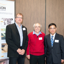
Above: Café Scientifique presenters (from left to right): Drs. Jeffrey Carson, Frank Prato (moderator), Ting-Yim Lee and Udunna Anazodo.
An image of the future: Innovations in imaging research
11:00pm - 1:00am
Lawson Health Research Institute has long been a leader in biomedical imaging. The first MRI images in Canada were captured at St. Joseph’s Hospital and we were the first in the country to install PET/CT and PET/MRI scanners. New developments in imaging research continue to enhance the diagnosis, prevention and treatment of a wide range of diseases, from cancer to PTSD.
You are invited to Lawson’s Café Scientifique, a free community event providing an informal opportunity to get involved with science. Hear a panel of Lawson Imaging scientists discuss their cutting-edge work and have the opportunity ask questions as part of an open-forum discussion to gain insights from the speakers, and from one another.
In celebration of Canada’s 150th anniversary as a nation, this event is the first of a two-part series focusing on the future vision for health care in our country and the legacy our research will leave.
Presented Talks
- “Imaging of the heart: Seeing the cause of chest pain more clearly”
Dr. Ting-Yim Lee - “Using light and sound to improve breast surgery”
Dr. Jeff Carson - “How imaging can improve the management of epilepsy”
Dr. Udunna Anazodo - MODERATOR – Dr. Frank Prato
Registration
To register, please complete our registration form. To sign up to our email list for notification of future events, please email @email.
Event Information
Date: Tuesday, May 23, 2017
Time: 7 to 9 p.m.
Location: Mercato at Brescia University College, Clare Hall, 271 Ramsay Road, London ON, N6G 0S2
… 2000-2001 June 2001 Dr. Lauren McCurdy Director of Breast Imaging, St. Joseph’s Health Care, London Associate … Patsy Cline, and raises funds for St. Joseph’s Hospital’s Breast Imaging Program Mitchell and Kathryn Baran Family, … in support of St. Joseph’s Diagnostic Imaging for breast health Dyer Brown hosts its annual golf tournament …
Annual Report to the Community 2000-2001 June 2001 Dr. Lauren McCurdy Director of Breast Imaging, St. Josephs Health Care, London Associate Professor, UWO Visit our web site: www.sjhc.london.on.ca For additional copies of the report, complete financial statements, salary disclosure information or ot...
… 6 WAIT TIMES CUT FOR WOMEN FACING BREAST CANCER … the patient experience and advancing key roles in breast care, complex chronic disease management, and … or somebody in the future." WAIT TIMES CUT FOR WOMEN FACING BREAST CANCER In less than two years, the Breast Care Centre …
Annual Report 2012-2013 Welcome to the 2012-2013 Annual Report Contents Annual Report 2012-2013 ......................................................................................................................... 1 Welcome to the 2012-2013 Annual Report ............................................
… 7 ENHANCING CARE FOR BREAST CARE PATIENTS … well, and the CCAC was very supportive.” ENHANCING CARE FOR BREAST CARE PATIENTS It’s a designer gown that will never … kind hospital gown haute couture in the Norton Lucille Wolf Breast Care Centre. Members of the Breast Care Program team …
Annual Report 2013-2014 Welcome to our 2013-2014 Annual Report Contents Annual Report 2013-2014 ............................................................................................................................. 1 Welcome to our 2013-2014 Annual Report ........................................
… 15, women from across the region attended the 2104 BRA (Breast Reconstruction Awareness) Day at St. Joseph’s Hospital in London to learn about breast reconstruction post mastectomy. They heard directly … group for women by women who have had or are considering breast reconstruction. They shared their experiences and …
Annual Report 2014-2015 Welcome to the 2014-2015 Annual Report Contents Annual Report 2014-2015 ............................................................................................................................. 1 Welcome to the 2014-2015 Annual Report ........................................
… Revolutionary new breast imaging technology comes to St. Joseph’s … and the completion of $3.0 million of investments in our Breast Care program, renewing five mammography units. With … has the ability to perform contrast-enhanced mammograms and breast tomosysthesis (3D mammography) for our patients. …
Annual Report 2017-2018 Welcome to the 2017-2018 Annual Report Contents Annual Report 2017-2018 .......................................................................................................... 1 Welcome to the 2017-2018 Annual Report ...........................................................
… Care Visits 34,791 • Allergy and Immunology Program • Breast Care Program • Cardiac Rehabilitation and Secondary … Care Visits 34,791 • Allergy and Immunology Program • Breast Care Program • Cardiac Rehabilitation and Secondary …
St. Josephs Health Care London Renowned for compassionate care, St. Josephs is one of the best academic health care organizations in Canada dedicated to helping people live to their fullest by minimizing the effects of injury, disease and disability. FACTS & FIGURES Staff 3,970 Physicians* 1,095 Res...
… cataract and retinal surgery. St. Joseph’s also focuses on breast cancer, ear, nose and throat, gynecological, dental … ultrasound screen closely, it was obvious the lump in her breast had changed. Two years ago, it wasn’t a threat. Now, … about mammography screening. Her sister died at age 53 of breast cancer. When a tiny, non-palpable lump was discovered …
For additional copies of this publication, or other information about St. Josephs Health Care, London or St. Josephs Health Care Foundation, please call 519-646-6034. St. JoSephS Because of St. Josephs... Care and research across our region our people I love my work our SucceSSeS My baby is alive Su...
Beneficial bacteria may protect against breast cancer
Dr. Gregor Reid, a scientist at Lawson Health Research Institute, and his Western University PhD student, Camilla Urbaniak, have previously shown that live bacteria are present in the breast tissues of healthy women. This proves the existence of a breast tissue microbiome. In past studies, Reid and Urbaniak have also proven that human milk contains beneficial bacteria. “Since breastfeeding decreases a woman’s risk for breast cancer, we wondered if beneficial bacteria, like those found in human milk, may be playing a role in lowering the risk of cancer and whether other types of bacteria could be influencing cancer formation,” said Dr. Reid.
To explore these questions, Urbaniak obtained breast tissue samples from 58 women who had either benign or cancerous tumours. In addition, she obtained 23 samples from healthy women undergoing breast reductions or enhancements. Through an analysis of these tissues, Urbaniak found that the bacteria present in the breasts of healthy women differ from those found in the breasts of women with breast cancer.
Women with breast cancer had elevated levels of both Escherichia coli (E. coli) and Staphylococcus epidermidis. A NASA study has confirmed the London research findings and further identified bacteria associated with breast cancer. Urbaniak and Reid went even further by showing that these bacteria can cause significant damage, known as double-stranded breaks, to DNA. When this occurs, the body tries to repair the damage. However, these repairs often result in errors which can lead to the development of cancer.
Tissues taken from the breasts of healthy women showed high levels of Lactobacillus and Streptococcus, known to promote health and display characteristics that can prevent cancer. For example, Streptococcus produces antioxidants that can help prevent DNA damage.
“This study provides clear evidence that the breast tissue microbiome differs between healthy women and those with breast cancer,” said Dr. Reid. “Our colleagues in Spain have recently shown that probiotics with lactobacilli can be ingested by women and reach the mammary gland. This raises the questions of whether women, especially those at risk for breast cancer, should take probiotics to increase the proportion of beneficial bacteria in the breast.”
In addition to prevention, this finding could have potential for helping with the management of patient disease. “It may be possible to increase the abundance of beneficial bacteria at the expense of harmful ones through the use of probiotics,” said Dr. Reid. “Antibiotics targeting harmful bacteria may also be another option for improving breast cancer management. Additional research is warranted to further explore the role of the breast tissue microbiome in the development and prevention of breast cancer.”
“In the near future, I hope we can discover which bacteria or combination of bacteria promote cancer development and which ones could help protect against it, and the mechanisms by which they do so,” said Urbaniak. “The next logical step would then be to unravel how we can use this knowledge to protect women from getting breast cancer or how we can manipulate the microbiome to help treat cancer once a woman gets it.”
The study, “The microbiota of breast tissue and its association with breast cancer”, was featured in Applied and Environmental Microbiology, a journal of the American Society for Microbiology. Dr. Gregor Reid is the Director, Canadian Centre for Human Microbiome and Probiotic Research at Lawson Health Research Institute and a Professor of Surgery, and Microbiology and Immunology at Western University. Urbaniak, who recently completed her PhD at Western University, will soon be a postdoctoral fellow at the NASA Jet Propulsion Laboratory (JPL) in Pasadena, California where she will perform microbiome analyses on samples collected from astronauts as well as those collected from the surfaces of the International Space Station.
Scientist
… of $.5 billion by fall 2025/26. • Beginning Fall 2024, breast screening will begin at age of 40 yrs instead of 50 … of $.5 billion by fall 2025/26. • Beginning Fall 2024, breast screening will begin at age of 40 yrs instead of 50 …
P a g e | 1 Meeting of the Board of Directors Monday, November 27, 2023 3:30 pm start time St. Josephs Hospital Adams Boardroom A2-041 Via MS Teams video-conference MINUTES Minutes to be ratified next meeting Call to Order Nawaz Tahir The reflection was provided by Robert Raymond. Education Sessio...