Search
Search
308 Search Results:
… sites in the world piloting the next generation of diabetes management tools. há|Çz ÁzÉÉwË utvàxÜ|t àÉ y|z{à ÁutwË St. … |Ç ÑxÉÑÄxËá Ä|äxá Patients at an interdisciplinary pain management centre being developed at St. Joseph’s … left, is leading in the creation of an interdisciplinary pain management centre at St. Joseph’s Hospital where …
ELDON HAWKINS Mount Hope resident Eldon Hawkins, a former Parkwood Hospital spinal cord injury rehabilitation program and respite care patient, returns to Parkwood twice a week as a member of the Parkwood Fitness Centre, a fully accessible facility designed to assist adults with physical disabilitie...
… Regional Mental Health Care London), built in 1902, painted by London artist Deanna Ronson. Deanna has been a … Less than 24 hours later, she was home. At no time needing pain control, the avid walker was back in stride six days … and volunteers are as important as the work itself. To manage and minimize stress in the workplace, St. Joseph’s …
Annual Report 2008 | 2009 St. Josephs Health Care, London BECAUSE WE ALL NEED ST. JOSEPHS 1 THIS PAST YEAR HAS BEEN ONE OF MILESTONES AND MOMENTS. FROM THE MAJOR INITIATIVES, CHALLENGES AND ACHIEVEMENTS TO THE COUNTLESS ACTS OF CARE AND COMPASSION, WE MARK THEM ALL IN THIS, OUR ANNUAL REPORT TO THE ...
… crane EllisDon showed their support for St. Joseph’s by painting a 168 foot construction crane pink – making it the … Program at Parkwood Hospital. $6,262 to provide self-management resource kits for ongoing treatment for patients … depression, obsessive-compulsive disorder, chronic pain, and migraines. $3,145 to bring Christmas dinner to …
2013/2014 The Thoughtful Acts of a Giving Community One philanthropic gesture has the power to impact the lives of thousands. At St. Josephs Health Care Foundation, we are honoured to be the recipient of that philanthropy, making a difference to the health and well-being of our community and care t...
… in their lifetime, a condition that causes excruciating pain often compared to childbirth. The lithotripter, a … through socialization and provide diversion from chronic pain. Helping seniors and veterans maintain mobility Routine … Hospital, a new clinical research centre will address the management of complex chronic diseases such as diabetes, …
Community Stewardship Report 2014 2015 An average day at St. Josephs is filled with possibilities, whether its through innovative approaches to treatment or the potential offered by new medical technologies. Each day, these seemingly endless possibilities can be measured in the way people recovering...
2018 Scientist of the Year Award: Dr. Robert Teasell
Lawson scientist Dr. Robert Teasell is considered a global leader in neurorehabilitation research and has been instrumental in transforming clinical care in this area across Canada by ensuring that clinical practices are informed by the best available and up-to-date research evidence. In recognition of his accomplishments, he received the Scientist of the Year Award at the 2018 Lawson Impact Awards event this past spring.
Dr. Teasell leads the Collaboration of Rehabilitation Research Evidence (CORRE) research team at St. Joseph’s Health Care London’s Parkwood Institute. He is also the Medical Director of the Stroke Rehabilitation Unit at Parkwood Institute and has an active outpatient chronic pain practice.
He has led the development of three internationally renowned evidence-based reviews for stroke rehabilitation, brain injury and spinal cord injury, which are regarded as the three most comprehensive research syntheses in neurorehabilitation in the world. Dr. Teasell has advised and helped plan stroke care for all of Ontario’s 14 Local Health Integration Networks (LHINs) and six provincial healthcare systems. This is in addition to the many clinical guidelines and models of care he has helped develop and update.
Dr. Teasell also bridges the gap between research and clinical practice through collaborations between his research and clinical teams. His multidisciplinary Rehabilitation Knowledge to Action Project (REKAP) team received the 2014 Sandra Letton Quality Award for their quality improvement project designed to make Parkwood Institute a leader in stroke rehabilitation by improving care through implementation of best practices.
In addition to his neurorehabilitation research, Dr. Teasell has published extensively on chronic pain with a recent focus on the role of obsessive personality traits in determining chronic pain disability and coping abilities.
Drawing on his clinical and research expertise, Dr. Teasell has supervised many students and has been committed to developing the next generation of medical researchers.
“Dr. Teasell has been successful in a number of areas. Certainly in terms of publications and mentorship of students who have gone on and had very successful careers of their own. Despite a busy clinical schedule, he always makes a point of engaging with his research team every day. His staff and students really appreciate the opportunity to work with him,” says Dr. Cheryl Forchuk, Beryl and Richard Ivey Research Chair in Aging, Mental Health, Rehabilitation and Recovery, and Assistant Director, Lawson Health Research Institute.
Dr. Teasell has authored 335 peer-reviewed articles, as well as many other collaborative group peer-reviewed articles, book chapters, published abstracts, posters, presentations and monographs. He has also been the editor for 14 special journal editions and is on the editorial boards for Topics in Stroke Rehabilitation, Journal of Rehabilitation, and Pain Research and Management.
In 2016, he was invited to present the Ramon J. Hnatyshyn Lecture, the leading annual national stroke lecture at the Canadian Stroke Congress in Quebec City. In 2010, he received the Canadian Association of Physical Medicine and Rehabilitation Merit award for his many contributions to the field of physiatry. He was also awarded the Royal College of Physicians and Surgeons of Canada McLaughlin-Gallie Visiting Professor in 2012. This year he will be awarded the Post-Acute Stroke Award of Excellence from the American Congress of Rehabilitation Medicine and the National Stroke Association in the United States.
“I’ve received a lot of national and international awards but there’s nothing better than being recognized by your peers and particularly your peers in the city where you work. It’s been a nice acknowledgement of not just my work, but also the work by the whole research team and all the people who have supported me over the years,” says Dr. Teasell.
Scientist
… therapy (iCBT). This form of therapy empowers people to manage their mental health. With your support, Dr. Swati … symptoms related to their traumatic injury or chronic pain to iCBT. In The MacDonald/Franklin Operational Stress … Lisa. Creating the look of a neighbourhood on the unit took paint and props to bring it to life. But it wouldn’t have …
Back row left: Holly Bridge, Diane Davis-Miller, Barb Simmonds Front row left: Dr. Jennifer Bjazevic, Roger Blum, Leanne Summers, Dr. Hassan Razvi, Surgical Services You make health care innovation possible. Just one of the reasons why YOU HELPED ENHANCE OUR OPERATING ROOMS 20192020 Community Impact...
… years of service, and ensures the compliance with emergency management protocols. The Anne Toal & Paul Brisson Annual … to help people living with traumatic injury or chronic pain that have developed psychological symptoms as a result. … SJHCF Grants illnesses, substance abuse, and chronic pain associated with lifelong illnesses. Care teams had …
Page 1 of 13 2019-20 SJHCF Grants St. Josephs Health Care Foundation 2019-20 Grants PATIENT CARE (In Alphabetical Order) 3 Complex Cataract Surgery Instruments Trays, $23,122 - St. Joseph's Hospital Equipment used in cataract surgery for the Ivey Eye Institute. 10 Blanket Warmers, $43,180 - Mount Ho...
… active 62-year old from Cambridge works as an Elite Athlete Manager for True Hockey, selling equipment to NHL teams and … residents every day. ADVANCING UROLOGY SURGERY FOR REDUCED PAIN AND RECOVERY TIME The right tools in the right hands … tumours, this game-changing equipment is leading to less pain and shorter recovery times for patients. BREAKING DOWN …
St. Josephs Health Care Foundation 268 Grosvenor Street PO Box 5777 STN B London, ON N6A 4V2 519-646-6085 sjhc.london.on.ca/foundation CHARITABLE REGISTRATION NUMBER: BN 11918 3390 RR0001 facebook.com/stjosephslondon youtube.com/stjosephslondon twitter.com/stjosephslondon Connect with us YOUR SUPPOR...
2022 Media Releases
Researchers are combining new technologies to examine blood proteins in COVID-19 patients
Study aims to empower patients with Type 2 diabetes to take control of their health
Largest trial ever done in hemodialysis care examines optimal dialysis temperature
Virtual care associated with significant environmental and patient cost savings
London researchers discover novel method to diagnose long COVID
New study testing whether virtual groups can improve well-being in older adults
Leveraging virtual reality to manage pain in paediatric patients
London researchers adapt MRI technology to image salt within the kidneys
London researchers collaborating on national dementia prevention program
New tool shows promise in helping people manage traumatic brain injuries one pace at a time
Local researchers using artificial intelligence to lead the way in bedside lung imaging
New study aims to improve mental health treatments for stroke patients
Local scientists creates novel test that could easily diagnose repetitive blast injury
April 11, 2022
Study finds high percentage of patients with a severe COVID-19 infection will end up with kidney injury, often fatal
March 15, 2022
Study shows a decline in Veterans’ mental health throughout the pandemic
February 16, 2022
Shocking number of heart attack patients suffer dangerous hemorrhage following lifesaving treatment, study shows
February 10, 2022
Study examines new forms of treatment for those suffering from post-traumatic stress disorder
January 25, 2022
London ranks in top ten of Canada’s research hospitals
January 18, 2022
New study looking at advanced imaging to optimize treatments for prostate cancer patients
January 12, 2022
… 2025 Pain Management Rheumatology Workshop February - June …
… you are working with has called to talk to you about pain relief for a patient. You ask him to go ahead and order … has presented with fever, shakes/chills, fatigue, abdominal pain, and severe diarrhea. The patient is on contact … protecting her airway and will likely require more invasive management. You are uncomfortable with taking care of her …
My First Week of Residency: Survival Pearls & Call Resident Orientation: June 29 2018 Are you ready for a brand new chapter in your medical career? Here are some tips and tricks to survive your first few days, and CALL Welcome to Residency! You arrived in London yesterday for Orientation and you are...
… A guide to managing your pain during COVID-19 Our services Hospitals across Ontario … for the time being. What does this mean for the Pain Management Program? For now, our groups, workshops and … A guide to managing your pain during COVID-19 Our services Hospitals across Ontario …
A guide to managing your pain during COVID-19 Our services Hospitals across Ontario are doing everything they can to limit the spread of COVID-19, including rescheduling non- urgent outpatient clinic appointments for the time being. What does this mean for the Pain Management Program? For now, our g...
… Sleep Issues Following ABI April 5, 2011 15142663 Chronic Pain April 26, 2011 15142910 Survivor Stories 2011 May 3, … April 12, 2016 54026579 Dealing with Headaches and Pain Following Brain Injury April 19, 2016 54027516 Brain … Sleep Issues Following ABI April 5, 2011 15142663 Chronic Pain April 26, 2011 15142910 Survivor Stories 2011 May 3, …
Revised March 16, 2016 How to Access Archived Webcasts of the Survivor & Family Education Series 1. Go to http://webcast.otn.ca 2. In the middle of the screen, under Archived Events, select Public. 3. In the Search Box in the top right hand corner, type: Parkwood ABI Survivor Series and press enter....
… • balance issues • vision issues • hearing changes • pain • weakness • seizures • feel tired all the time • … your memory and concentration • changes in your ability to manage yourself and your life • changes in your personality … • balance issues • vision issues • hearing changes • pain • weakness • seizures • feel tired all the time • …
CARING FOR THE BODY, MIND & SPIRIT SINCE 1869 Renowned for compassionate care, St. Josephs is one of the best academic health care organizations in Canada dedicated to helping people live to their fullest by minimizing the effects of injury, disease and disability through excellence in care, teachin...
… the two weeks some concrete, practical strategies to help manage issues such as light and noise sensitivity, headache, … approach. April 16 Dealing with Headaches and Pain Following ABI Speaker: Dr. Keith Sequeira Pain is often a common issue for people post brain injury …
Parkwood Institute Acquired Brain Injury Survivor and Family Education Series Spring 2019 Schedule The survivor and family education series is a forum open to individuals with acquired brain injury, their family and friends. Sessions will be Tuesday evenings from 6:30 to 8:00 pm. In London, the sess...
… may still develop osteoporosis Symptoms Osteoporosis is not painful unless you have fractures. Bone loss occurs without … the spine to collapse. This can result in height loss, pain and a deformed back. Diagnosis Several tests can detect … exposes the patient to a low level of radiation. Treatment Management of osteoporosis includes fall avoidance and …
Facts about Osteoporosis What is osteoporosis? Osteoporosis means porous or brittle bones. Weak bones can break easily; fractures of the hip, wrist and spine are commonly associated with osteoporosis. Bone is surprisingly dynamic. It is constantly remodelled; bits of bone are eaten away or resorbed,...
… Neuropathic Pain Seminar Saturday, February 8, 2020 0800-1600 St. … 1) Be able to diagnose various forms of neuropathic pain 2) Understand the diverse pathophysiology of … pain 3) Identify the optimal evidence based strategies for management of neuropathic pain 4) Identify when it is …
Neuropathic Pain Seminar Saturday, February 8, 2020 0800-1600 St. Josephs Hospital Shuttleworth Auditorium, Zone D, Level 0, Room D0-104 List Overall Learning Objectives: 1) Be able to diagnose various forms of neuropathic pain 2) Understand the diverse pathophysiology of neuropathic pain 3) Identi...
… is this medication prescribed? • Amitriptyline: o treats pain by increasing the concentrations of chemical messengers in the nervous system to reduce the pain messages arriving in the brain. o treats pain from … it may take a little while before you notice the pain management benefits of this medication. It may take about 4 …
June 16, 2020; Page 1 of 2 Amitriptyline (Elavil) Why is this medication prescribed? Amitriptyline: o treats pain by increasing the concentrations of chemical messengers in the nervous system to reduce the pain messages arriving in the brain. o treats pain from damaged nerves. The pain is usually d...
An image of the future: Innovations in imaging research
Lawson Health Research Institute (Lawson) has long been a leader in biomedical imaging. The first Canadian magnetic resonance imaging (MRI) of a human occurred at St. Joseph’s Health Care London (St. Joseph’s). The country’s first positron emission tomography/computed tomography (PET/CT) and positron emission tomography/magnetic resonance imaging (PET/MRI) scanners were also installed at St. Joseph’s. New developments in imaging research continue to enhance the diagnosis, prevention and treatment of a wide range of diseases, from cancer to post-traumatic stress disorder.
On May 23, Lawson hosted a Café Scientifique event where a panel of Lawson Imaging scientists discussed their cutting-edge work. Guests had the opportunity to ask questions as part of an open-forum discussion to gain insights from the speakers, and from one another.
In celebration of Canada’s 150th anniversary as a nation, this event is the first of a two-part series focusing on the future vision for health care in Canada and the legacy that research at Lawson will leave.
Imaging of the heart: Seeing the cause of chest pain more clearly
By Dr. Ting-Yim Lee, Lawson scientist, Medical Physicist at St. Joseph’s, professor at Western University’s Schulich School of Medicine & Dentistry, and scientist at Robarts Research Institute
When patients with chest pain arrive in the emergency department, they are given an electrocardiogram (ECG) and blood test. These diagnostic tests determine if the pain has a non-cardiac cause (such as heart burn), if it is caused by a heart attack, or if the patient has angina (plaque formation in the coronary arteries that either reduces or temporarily cuts off blood flow to the heart) but did not have a heart attack.
If a patient has angina, they are then given additional diagnostic testing to see whether a blood clot has formed and where it is located. This is determined by two different imaging techniques: x-ray imaging (angiogram) and nuclear imaging. This process is invasive and means that patients must be scheduled for two different exam days. Using two techniques also means that there can be image misalignment, and the images often provide poor detail.
Dr. Ting-Yim Lee’s lab has pioneered a Computed Tomography (CT) method for imaging blood flow to the heart muscle (CT Perfusion), which can help patients avoid unnecessary tests and treatment, as well as reduce health care costs.
“CT imaging is a non-invasive imaging technique that uses x-rays to create high-detail cross-sectional images of the body. Using this method, we can evaluate the degree of blockage in coronary arteries – with one diagnostic test instead of two,” says Dr. Lee.
Using light and sound to improve breast surgery
By Dr. Jeffrey Carson, Lawson scientist and associate professor at Western University’s Schulich School of Medicine & Dentistry
“Most women diagnosed with breast cancer undergo surgery, and months of chemotherapy and radiotherapy. They must deal with the discomfort, side-effects, emotional stress and financial burden of treatment. Almost one in four surgeries for breast cancer must be repeated, meaning many women have to go through this all over again,” says Dr. Jeffrey Carson.
In breast conserving surgery, there is a high chance of repeat surgery as the surgeon must see and remove 100 per cent of the tumour in order for it to be successful. They are not able to determine whether the entire tumour was removed until after the surgery has been completed.
Dr. Carson and his team at St. Joseph’s have developed a technology called Intraoperative Photoacoustic Tomography (iPAT), which has the potential to reduce the chance of repeat surgery for breast cancer. The technology is able to image surgery specimens in the operating room during surgery, allowing surgeons to determine whether the whole tumour has been removed before the surgery is complete.
How imaging can improve the management of epilepsy
By Dr. Udunna Anazodo, postdoctoral fellow at Lawson
Most patients with epilepsy are effectively treated with antiepileptic drugs. However, 36 per cent will not respond to the drugs. For these patients, surgery on the area of the brain that is causing seizures is the standard of care – if patients are good surgical candidates.
“If patients with epilepsy are to undergo surgery there must be a good indication of where the seizure focus is and it must be possible to determine that removing this portion of the brain will not affect brain function,” says Dr. Udunna Anazodo.
To see whether they are good candidates for surgery, patients must undergo an invasive procedure called intracranial monitoring, where electrodes are placed on the brain.
Dr. Anazodo has been studying how PET/MRI can be used to map seizures with the goal of minimizing the need for invasive intracranial monitoring. This technique makes it possible to locate areas in the brain that cause seizures and to see if the seizures affect brain functions.
See photos from the event on Lawson’s Facebook page.
Image
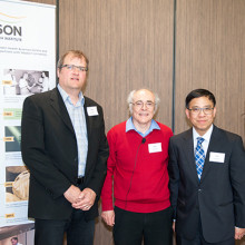
Above: Café Scientifique presenters (from left to right): Drs. Jeffrey Carson, Frank Prato (moderator), Ting-Yim Lee and Udunna Anazodo.
An image of the future: Innovations in imaging research
11:00pm - 1:00am
Lawson Health Research Institute has long been a leader in biomedical imaging. The first MRI images in Canada were captured at St. Joseph’s Hospital and we were the first in the country to install PET/CT and PET/MRI scanners. New developments in imaging research continue to enhance the diagnosis, prevention and treatment of a wide range of diseases, from cancer to PTSD.
You are invited to Lawson’s Café Scientifique, a free community event providing an informal opportunity to get involved with science. Hear a panel of Lawson Imaging scientists discuss their cutting-edge work and have the opportunity ask questions as part of an open-forum discussion to gain insights from the speakers, and from one another.
In celebration of Canada’s 150th anniversary as a nation, this event is the first of a two-part series focusing on the future vision for health care in our country and the legacy our research will leave.
Presented Talks
- “Imaging of the heart: Seeing the cause of chest pain more clearly”
Dr. Ting-Yim Lee - “Using light and sound to improve breast surgery”
Dr. Jeff Carson - “How imaging can improve the management of epilepsy”
Dr. Udunna Anazodo - MODERATOR – Dr. Frank Prato
Registration
To register, please complete our registration form. To sign up to our email list for notification of future events, please email @email.
Event Information
Date: Tuesday, May 23, 2017
Time: 7 to 9 p.m.
Location: Mercato at Brescia University College, Clare Hall, 271 Ramsay Road, London ON, N6G 0S2