Search
Search
2194 Search Results:
… Feb 2024.doc Complex Care Program – Parkwood Institute Patients Eligible for transfer to the Long Term Ventilation … will best be met in Complex Care at Parkwood Institute. Patients are expected to meet the following criteria: 1. … breathing, the more the better; however will accept patients with no spontaneous breathing capability. • …
Critieria for admission to LTV Program Feb 2024.doc Complex Care Program Parkwood Institute Patients Eligible for transfer to the Long Term Ventilation Beds There are six beds within the Complex Care Program at Parkwood Institute that provide a clinical setting for individuals requiring long-term m...
… that the patient will return home Admission Guidelines: Patients are admitted for pain and symptom management, or … that the patient will return home Admission Guidelines: Patients are admitted for pain and symptom management, or …
How to refer a patient to Palliative Care Palliative Care provides pain management and relief of symptoms to those experiencing life-threatening, progressive or terminal illness. Palliative Care focuses on physical, psychological, social and spiritual needs while remaining sensitive to the uniquenes...
H-SAA Amending Agreement Extension to March 31, 2017 Page 1 H-SAA AMENDING AGREEMENT THIS AMENDING AGREEMENT (the Agreement) is made as of the 1st day of October, 2016 B E T W E E N: SOUTH WEST LOCAL HEALTH INTEGRATION NETWORK (the LHIN) AND St. Joseph's Health Care, London (the Hospital) WHEREAS t...
Hybrid PET/MRI Symposium
12:15pm - 10:00pm
On February 12, 2012, the first hybrid PET/MRI in Canada, and fifth in the world, was installed in London, Ontario at St. Joseph's Hospital. In 2016, hybrid PET/MRI started up in Toronto (UHN) and Ottawa (Royal) with new sites committed in Toronto (Sunnybrook), Edmonton, Winnipeg and Vancouver.
This one day symposium will summarize the London experience over the last five years. This will include the technological challenges that have been met (e.g. the need for PET compatible RF coils, MRI directed attenuation correction in brain imaging and the advantage of co-siting a medical cyclotron and radio-chemistry facility) and those that remain (e.g. reimbursement for routine patient use, hybrid reconstruction, hybrid kinetic modeling and respiratory and cardiac corrections for true registration). Experience with applications in cardiology, epilepsy, prostate cancer and rectal cancer will be presented. Presenters from Ottawa and Toronto will also look to the future of hybrid PET/MRI in cardiology, neurodegeneration and psychiatry.
Researchers and students interested in hybrid PET/MRI and its potential role in clinical diagnosis will benefit from this symposium. This includes those interested in technology development and those interested in using PET/MRI to discover the mechanisms of disease and the assessment of therapies. In one full day, attendees will gain knowledge concerning the strengths and challenges of hybrid PET/MRI technology and its growing role in oncology, neurology and cardiology. With a growing number of sites in Canada, there will be an excellent opportunity to form collaborations and the opportunity to establish a Canadian network.
Date and Time: Tuesday, March 14, 2017, 8:15 a.m. to 5:45 p.m.
Location: St. Joseph's Hospital, Shuttleworth Auditorium (D0-104)
Schedule: View the event schedule.
Registration: Online registration is now closed. Please contact Shelagh Ross at @email by Friday, March 10 to register.
ICU patients with non-brain-related injuries may suffer undetected cognitive dysfunction
LONDON, ON - A new study led by Western University and Lawson Health Research Institute has found that most patients entering hospital intensive care units (ICU) for non-brain-related injuries or ailments also suffer from some level of related cognitive dysfunction that currently goes undetected in most cases.
The findings were published today in the influential scientific journal, PLOS One.
Many patients spend time in the ICU for reasons that have nothing to do with a known brain injury, and most health care providers and caregivers don’t have any evidence to believe there is an issue with the brain. For example, a patient may have had a traumatic injury that does not involve the brain, yet still requires breathing support to enable surgeons to fix damaged organs, they may have issues with their heart or lungs, they may contract a serious infection, or they may simply be recovering from a surgical procedure like an organ transplant that has nothing directly to do with their brain.
For the study, Western researchers from the Schulich School of Medicine & Dentistry and the Brain and Mind Institute and researchers from Lawson assessed 20 such patients as they left the ICU and every single patient had detectible cognitive deficits in two or more cognitive areas of investigation, including memory, attention, decision-making and reasoning. Again, this is in spite of the fact that, on the face of it, they had no clear brain injury.
The discovery was made using online tests, developed by renowned Western neuroscientist Adrian Owen and his teams at the Brain and Mind Institute and BrainsCAN, which were originally designed to examine cognitive ability in patients following brain injuries but for this purpose, are being used to detect cognitive deficits in people who have spent time in an intensive care unit without a diagnosed brain injury.
“Many people spend time in an intensive care unit following a brain injury and, of course, they often experience deficits in memory, attention, decision-making and other cognitive functions as a result,” explains Owen, a professor at Schulich Medicine & Dentistry. “In this study, we were interested to see how patients without a specific brain injury fair after leaving the ICU. The results were astonishing.”
Why cognitive ability declines even in non-brain related visits to the ICU likely varies from patient to patient, but Dr. Kimia Honarmand from Schulich Medicine & Dentistry says the lesson to be learned is that many conditions affect brain function, even though they might not directly involve the brain.
“If you are having trouble breathing, your brain may be starved of oxygen. If you have a serious infection, the inflammation that occurs as a result of infection may affect brain function. If you are undergoing major surgery, you might be given drugs and have procedures that may affect your breathing, which in turn may affect the flow of oxygen to the brain,” explains Dr. Honarmand. “What we have shown here is that all or any of these events can lead to deficits in brain function that manifest as impairments in cognition. And healthy cognition is a vital determinant of functional recovery.”
Dr. Marat Slessarev, Lawson Scientist, says these findings can shift how the medical community treats incoming patients and more importantly, outpatients following ICU visits.
“Historically, the clinical focus has been on just survival. But now we can begin to focus on good survival,” says Dr. Slessarev, also an associate member at the Brain and Mind Institute and an assistant professor at Schulich Medicine & Dentistry. “These sensitive tests will enable doctors to both detect cognitive impairment and track cognitive performance over time, which is the first step in developing processes for optimizing brain recovery.”
-30-
About Western
Western delivers an academic experience second to none. Since 1878, The Western Experience has combined academic excellence with life-long opportunities for intellectual, social and cultural growth in order to better serve our communities. Our research excellence expands knowledge and drives discovery with real-world application. Western attracts individuals with a broad worldview, seeking to study, influence and lead in the international community.
About The Schulich School of Medicine & Dentistry
The Schulich School of Medicine & Dentistry at Western University is one of Canada’s preeminent medical and dental schools. Established in 1881, it was one of the founding schools of Western University and is known for being the birthplace of family medicine in Canada. For more than 130 years, the School has demonstrated a commitment to academic excellence and a passion for scientific discovery.
About Lawson Health Research Institute
Lawson Health Research Institute is one of Canada’s top hospital-based research institutes, tackling the most pressing challenges in health care. As the research institute of London Health Sciences Centre and St. Joseph’s Health Care London, our innovation happens where care is delivered. Lawson research teams are at the leading-edge of science with the goal of improving health and the delivery of care for patients. Working in partnership with Western University, our researchers are encouraged to pursue their curiosity, collaborate often and share their discoveries widely. Research conducted through Lawson makes a difference in the lives of patients, families and communities around the world. To learn more, visit www.lawsonresearch.ca.
Media Contacts
Celine Zadorsky
Senior Media Relations Consultant
Communications & Public Engagement
T: 519-685-8500 ext. 73502
Celine.zadorsky@lhsc.on.ca
Illuminating the body's smallest secrets
Lawson Research Institute’s cyclotron facility is a formidable partner in the fight against disease.
Tucked behind the protective lower walls of St. Joseph’s Hospital lies a futuristic workshop of sorts – a place where science, technology and leading-edge medical care converge.
Within a behemoth, 62-ton machine, a swirling vortex of powerful magnetic fields and electric pulses is creating bursts of radioactive isotopes – tiny, potent sparks of life-saving potential. In the hands of technologists, researchers and clinicians, these chemical elements become diagnostic tracers and therapeutic agents, each particle revealing secrets of the human body.
The machine is a cyclotron - a type of particle accelerator and the only one of its kind in the region. At Lawson Research Institute’s Cyclotron & PET Radiochemistry Facility, scientists are producing a steady and timely supply of short-lived radioisotopes every day to study, detect and treat disease.

These radioisotopes become a beacon in positron emission tomography (PET) scans, illuminating the hidden shadows of cancer and other diseases. Others provide a precise map of the intricate pathways of blood flow, biological functions, location of specific cells and proteins, and the body’s skeletal architecture.
A formidable partner in the fight against disease, “the cyclotron facility is a hub for Southwestern Ontario that is uncovering the possibilities for improving patient care in numerous ways,” says Michael Kovacs, PhD, Lead, Lawson’s Cyclotron & PET Radiochemistry Facility and Program Lead, Lawson Imaging Research Program.
St. Joseph’s cyclotron supports a wide variety of research projects including imaging applied to oncology, cardiology, neurology, psychiatry, metabolic disease, infectious diseases, bioelectromagnetics and other areas.
“The scope of discoveries already making a difference, and the possibilities within reach, are a source of great pride for Lawson and for London,” adds Frank Prato, PhD Lawson scientist and Chief Medical Physicist at St. Joseph’s.
For Kovacs, Prato, their teams and partners, St. Joseph’s cyclotron is a testament to ingenuity and innovation, a world where every spin and burst of charged particles brings a promise of hope and healing.
Powering innovation
Generous donors to St. Joseph’s Health Care London have made both advanced research and next-level technology a reality. During the past few years, more than $1.1 million in donations funded extensive renovations to the Cyclotron & PET Radiochemistry Facility, making it possible to increase production of isotopes and expand life-saving care.
Recently, $1 million in donations supported a new PET/CT – the heart of the Canada’s first national GE centre of excellence in molecular imaging and theranostics being developed at St. Joseph’s Hospital.
Imaging
Lawson is in the international forefront of medical imaging
Research that takes a deep look inside the human body and seeks to understand how tissues, bones, organs and cells work, and then guides next steps when the body doesn’t work as it should.
At Lawson, we see things more clearly than anyone.
Lawson leads medical and imaging research. Learn more:
We’re home to a host of Canadian and world firsts that have resulted in new pathways of diagnosis, treatment and care.
We’re recognized as pioneers in molecular imaging: a virtual alphabet of MRIs, PET scans, CT scans and more.
With technologies that also include our state-of-the-art cyclotron, we keep pushing the boundaries of these technologies so that soon we hope to test treatments with the same pinpoint precision and technology we use for imaging. This is a new and exciting field called theranostics, and it offers the very real possibility of a magic bullet to eradicate some cancers.
Our award-winning scientists, trainees and staff share their discoveries to improve patient lives locally and around the world.
Lawson is a global leader in imaging research. Read on to see where it can lead you.
Lawson Intranet (password protected) | ICES: data-focused research | Animal Care Committee
Imaging “hidden” regions of the heart
After suffering a heart attack, some patients develop a microvascular obstruction, an area of the injured heart with extremely poor blood flow. These patients are at an increased risk of developing heart failure in the future.
Medical imaging technologies such as magnetic resonance imaging (MRI) and positron emission tomography (PET) can be used to study the remodeling process after a heart attack that can lead to a microvascular obstruction. However, poor blood flow makes it difficult to get contrast agents into the obstruction. Contrast agents are used in medical imaging to show contrast between different types of tissue, such as damaged and healthy tissue.
Benjamin Wilk, a PhD candidate at Lawson Health Research Institute and Western University’s Schulich School of Medicine & Dentistry, will investigate whether a hybrid PET/MRI system and a new method of administering contrast agents can allow researchers to image microvascular obstructions and study these “hidden” regions in the heart.
Contrast agents are usually injected as a bolus, meaning the entire injection is administered immediately. In this study, participants will instead receive a constant infusion of an MRI contrast agent and PET tracer, which means the injection will be delivered over the course of an hour. The MRI contrast agent they are using is sensitive to blood flow and scar tissue, and the PET tracer is sensitive to inflammatory cells.
This will allow researchers to study the anatomy, blood flow and inflammatory processes in microvascular obstructions a week after heart attack. Participants will then be imaged again after six weeks to study the long-term effects on heart function.
“Studying the heart after a heart attack using novel contrast agent injection strategies with simultaneous PET/MRI could provide crucial information for treatment planning, helping us reduce the number of people affected by heart failure in the future,” says Wilk. “This project could also lead to further research into finding better ways to administer PET tracers and MRI contrast agents. These methods could be applied to different diseases as well.”
Wilk received a Lawson Internal Research Fund (IRF) Studentship to conduct the study, which will be supervised by Dr. Frank Prato, Assistant Director, Lawson and leader of the Lawson Imaging research program at St. Joseph’s Health Care London.
“Lawson's IRF is valuable for students for many reasons. It not only allows us to conduct further research, it also enriches our experience by giving us opportunities to write grants and attend conferences,” adds Wilk.
The IRF is designed to provide Lawson scientists the opportunity to obtain start-up funds for new projects with the potential to obtain larger funding, be published in a high-impact journal, or provide a clinical benefit to patients. Funding is provided by the clinical departments of London Health Sciences Centre and St. Joseph’s Health Care London, as well as the hospital foundations (London Health Sciences Foundation and St. Joseph’s Health Care Foundation).
Scientist
Imaging the microbiome
Normally samples of bacteria must be removed from their microbiome environment for analysis, which can lead to changes in their metabolic activity and other behaviors, hindering our ability to accurately study the gut or urogenital microbiome.
“This could be avoided if we are able to observe the bacteria in the body using Magnetic Resonance Imaging (MRI),” says Sarah Donnelly, MSc student at Lawson Health Research Institute and the Department of Microbiology and Immunology and collaborative Molecular Imaging program at Western University’s Schulich School of Medicine & Dentistry.
She is investigating the possibility of using magnetically-labelled bacteria with MRI to more directly study microbial interactions in urological and other conditions.
“The hope is that in the future we can use imaging technologies to visualize aspects of the microbiome in its healthy state compared to diseased states to see the early signs of disease and take preventative measures or allow for early intervention,” she says.
Donnelly has received a Lawson Internal Research Fund (IRF) Studentship to conduct the study, which will be supervised by Dr. Jeremy Burton, scientist in Lawson’s Human Microbiome and Probiotics research program at St. Joseph’s Health Care London (St. Joseph’s) and appointed to the Departments of Surgery and Microbiology & Immunology at Schulich Medicine & Dentistry; and Dr. Donna Goldhawk, scientist in Lawson’s Imaging research program at St. Joseph’s and assistant professor in the Department of Medical Biophysics at Schulich Medicine & Dentistry.
Escherichia coli (E. coli) are a common bacterium found in the human gut microbiome and frequently cause non-intestinal conditions like urinary tract infections. The researchers will program E. coli to express an iron uptake gene, magA. This gene is taken from another type of bacteria called magnetotactic because of their response to Earth’s magnetic field. The researchers will study whether the increase in iron uptake caused by magA expression will allow MRI to detect the magnetic signal more clearly than it would in images of untransformed E.coli. This would make it possible to see the bacteria’s behavior in living subjects without removing the bacteria cells from the microbiome environment.
They will then use this technique to study how magA labelled bacteria affect biofilm on medical devices. A biofilm is a structure produced when certain bacteria adhere to a surface and then stick together.
They will also analyze how lithotripsy affects the bacteria’s spatial distribution and interactions in three-dimensional models of kidney stones. Lithotripsy uses shockwaves to break up kidney stones into smaller pieces that are able to pass naturally out of the body. However, these shockwaves not only affect kidney stones. The waves are sent throughout the tissue, and the bacteria living on these tissues may also be affected.
“While lithotripsy is effective in treating kidney stones, we don’t know the side effects of lithotripsy on the microbiome. The shockwaves could disturb the bacteria, potentially leading to diseases caused by an imbalance between helpful and harmful bacteria,” says Donnelly.
These laboratory models will allow the researchers to perform studies in vivo (in animal models) in the future.
“Health research is very important for the development of new technologies and treatments but it is often difficult to secure funding. The IRF program allows students to pursue research that would not otherwise be possible,” explains Donnelly.
The IRF is designed to provide Lawson scientists and students the opportunity to obtain start-up funds for new projects with the potential to obtain larger funding, be published in a high-impact journal, or provide a clinical benefit to patients. Funding is provided by the clinical departments of London Health Sciences Centre and St. Joseph’s Health Care London, as well as the hospital foundations (London Health Sciences Foundation and St. Joseph’s Health Care Foundation).
Scientist
Improved imaging for prostate cancer
Prostate cancer is the most commonly diagnosed cancer among Canadian men. However, with improved testing and better treatment options, the number of deaths from the disease has been declining over the last several years.
Scientists at Lawson Health Research Institute are working to continue this trend by testing improved prostate cancer imaging using a new molecule. Known as a Prostate Specific Membrane Antigen (PSMA) probe, the new molecule is used in Positron Emissions Tomography (PET) scans. The probe targets PSMA, a unique molecule on prostate cancer cells, to provide highly specific images for better diagnosis and management of patient disease.
PET probes are used in imaging to correctly diagnose cancer. The probes are injected into a patient where they spread to identify sites of disease. The most common PET probes are suitable for many types of cancer, but are not as sensitive in identifying prostate cancer. PSMA probes provide higher accuracy by targeting PSMA molecules, which are highly over-expressed on prostate cancer cells.
PSMA probes are gaining popularity across the globe. This specific probe is a molecule called 18F-DCFPyL and was developed by Dr. Martin Pomper at the John Hopkins Hospital in Baltimore. Dr. Pomper, also a Scientific Advisor to Lawson’s prostate imaging team, worked in collaboration with Canada’s Centre for Probe Development and Commercialization (CPDC) to bring the probe to our nation.
Lawson’s Canadian Institutes of Health Research (CIHR) Team in Image Guidance for Prostate Cancer gained early access to the PSMA probe due to a history of close collaboration with Dr. Pomper and the CPDC. Marking the first time a PSMA probe has been used in Canada, the team captured PET/MRI and PET/CT images from a 64-year-old prostate cancer patient on March 18, 2016 at St. Joseph’s Hospital.
“This is a tremendous step forward in the management of prostate cancer,” said Dr. Glenn Bauman, a Lawson scientist and Radiation Oncologist at London Health Sciences Centre. “PSMA probes have the potential to provide increased accuracy and detection which leads to better, personalized treatment.”
Lawson plans to study the probe with an additional 20 men over the next two years as part of an ongoing clinical trial funded by the Ontario Institute for Cancer Research (OICR). Lawson scientists are working with researchers across Ontario to develop other clinical trial protocols that will use 18F-DCFPyL to measure responses to drug treatments and to evaluate men with suspected recurrence of prostate cancer after radiotherapy.
“The goal of these studies it to establish the value of PSMA probes, particularly18F-DCFPyL, and provide evidence to support the use of these probes in routine clinical care,” said Dr. Bauman.
Donor funding through London Health Sciences Foundation was one catalyst in this research, providing initial funding to hire Research Associate, Catherine Hildebrand, who set up citywide cancer imaging workshops and helped the team prepare successful grant applications to secure key funding from CIHR and OICR.
Improving mental health treatments for stroke patients is the focus of a new study
LONDON, ON- Strokes affect approximately 400,000 Canadians each year and can be debilitating. They can negatively affect a person’s cognition and mobility, and severely impact mental health and wellbeing.
A team at Lawson Health Research Institute are looking to improve mental health treatments and resources for patients who have experienced a stroke. The team will recruit 100 stroke patients to assess whether the completion of a guided therapy program can improve mental health and quality of life.
“More than sixty per cent of patients experience depression after stroke,” says Dr. Robert Teasell, Lawson Scientist and Physiatrist at St. Joseph’s Health Care London’s Parkwood Institute. “Having a stroke itself makes people more vulnerable and makes people feel their lives have changed negatively.”
During stroke rehabilitation, patients are typically offered mental health treatments, but the research team say it is post rehabilitation that stroke patients tend to experience worsening depression.
“Publicly funded allied health care services are available at inpatient and outpatient care; however, psychology is often limited across the rehabilitation continuum from acute to community care,” says Dr. Swati Mehta, Lawson Scientist. “We are looking at how we can provide a program that is cost effective to help those who have these barriers to access mental health services.”
“Patients have described to me that they feel like they have been dropped off a cliff because of the lack of resources once their programs have ended,” adds Dr. Teasell.
The study will examine the use of cognitive behavioral therapy (CBT), which is an evidence based psychological intervention that aims to provide people with increased coping ability and self-efficacy. Participants will complete a 10-week guided program with specific targeted lessons, tailored to the needs of those post stroke, delivered virtually through a trained clinician. They will then complete a questionnaire to see if there have been any improvements to self-efficacy and emotional wellbeing.
“We have found this form of therapy (CBT) has been very effective and feasible for spinal cord injury patients with mild traumatic brain injury and we want to see how a modified version could potentially help those with stroke and depression,” says Randy Upper, Clinical Research Associate at Lawson.
If CBT is proven effective through this study, Dr. Mehta hopes it will encourage similar programming that would be available to stroke patients after rehab.
“We are hoping we can connect with community organizations and work with them to implement this program in a service delivery model that would be easily accessible for stroke patients living in the community.”
Recruitment for this study is currently underway, those interested in taking part can email Dr. Swati Mehta at: @email
Scientist
Media Contact
Celine Zadorsky
Senior Media Relations Consultant
Communications & Public Engagement
T: 519-685-8500 ext. 73502
Celine.zadorsky@lhsc.on.ca
Improving palliative cancer treatment with existing diagnostic scans: Study reveals promising results
A recent study from London Health Sciences Centre and Lawson Health Research Institute suggests that using existing diagnostic CT scans in planning simple palliative radiation treatments can significantly cut down the waiting time for urgent treatment, resulting in a better experience for cancer patients.
“Reducing the time patients spend in a cancer centre has far-reaching benefits,” said lead study author Melissa O’Neil an Advanced Practice Radiation Therapist at London Health Sciences Centre’s (LHSC) London Regional Cancer Program (LRCP). “Faster treatment initiation means quicker relief from symptoms for patients. Utilizing existing scans is also cost-effective and frees up appointment slots or staff, allowing us to accommodate and assist more patients in need.”
Palliative radiation therapy is used to relieve symptoms in patients whose cancers cannot be cured. It’s often used when tumours cause pain, neurological issues or breathing problems such as blocked airways.
In the current standard practice, patients referred for palliative radiation typically require a CT simulation scan before starting their treatment. This scan creates 3D images that the patient's health care team uses to develop a customized radiation treatment plan. Unfortunately, this process often takes several hours, even with efforts to speed it up.
However, many of these patients have undergone previous diagnostic CT scans as part of their routine medical care. Previous research has shown that radiation oncology teams can create suitable palliative treatment plans for patients with bone and soft tissue metastases using these existing scans. This approach is less time-consuming than the more intensive simulation scans.
In the current study, O’Neil and her colleagues explored whether using existing CT scans to plan treatment before a patient arrives at the cancer centre could reduce their wait time while still ensuring appropriate care. They randomly assigned 33 patients who needed palliative radiation for tumours in their chest, abdomen or pelvis to either the standard treatment planning with on-site CT simulation scans or to treatment planning using diagnostic CT scans taken within the previous 28 days.
The study found that patients who didn't need the extra CT simulation scan spent much less time at the cancer centre on the day of their treatment – just under 30 minutes compared to nearly five hours for the others. Treatments were delivered successfully, and patient perception on time spent at the cancer centre was improved for those whose treatment planning used diagnostic CT scans taken without the previous 28 days.
"For patients who need radiation to help treat symptoms of cancer, it's important for us to get them treated quickly and to minimize the time they spend waiting for medical appointments,” said Dr. David Palma, Radiation Oncologist at LHSC and Associate Scientist at Lawson. “This trial shows that this new approach not only saves resources by reducing the number of scans we do, but also substantially reduces the time patients spend waiting for urgent radiation.”
"These findings are incredibly promising, especially in light of the nationwide shortage of radiation therapists," said Dr. Michael Ott, Physician Department Executive for Oncology at LHSC. “Work like this has benefits that can reach far beyond London, offering more relief for patients across the country."
The findings were presented at the American Society for Radiation Oncology’s Annual Meeting on Oct. 3, 2023. This meeting is recognized globally as the leading radiation oncology scientific event, drawing more than 8,500 attendees each year.
While the study shows promise, the research team said it's important to note that using prior diagnostic scans may not be suitable for every type of cancer or patient. It depends on the specific area being treated and the technique used.
For more information, please contact:
Jessica Rabaey
Communications Consultant
London Health Sciences Centre
T: 519-685-8500 ext. 77728
Jessica.rabaey@lhsc.on.ca
About Lawson Health Research Institute
Lawson Health Research Institute is one of Canada’s top hospital-based research institutes, tackling the most pressing challenges in health care. As the research institute of London Health Sciences Centre and St. Joseph’s Health Care London, our innovation happens where care is delivered. Lawson research teams are at the leading-edge of science with the goal of improving health and the delivery of care for patients. Working in partnership with Western University, our researchers are encouraged to pursue their curiosity, collaborate often and share their discoveries widely. Research conducted through Lawson makes a difference in the lives of patients, families and communities around the world. To learn more, visit www.lawsonresearch.ca.
Improving recovery and rehabilitation for patients with mental illness
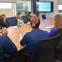
Over 130 hospital-based clinical, administrative and research staff members, persons with lived experience of mental illness, family caregivers, peer and community supporters, and staff from local community mental health agencies attended the 18th Annual Mental Health Research & Innovation Half Day on November 1, 2017. The event provided an opportunity to learn more about mental health research at Parkwood Institute and the Southwest Centre for Forensic Mental Health Care (Southwest Centre), part of the St. Joseph’s Health Care London (St. Joseph’s) family.
“This year’s Mental Health Research and Innovation Half Day was one of the best attended in our history of hosting the event. We had a very diverse and engaged audience with great energy and a lot of enthusiasm,” says Dr. Arlene MacDougall, Director of Research and Innovation for mental health care at St. Joseph’s and Lawson Health Research Institute (Lawson).
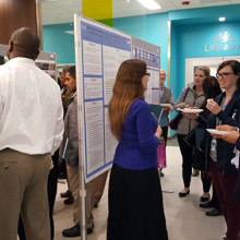
Exciting recent projects were showcased though talks highlighting excellence in recovery and rehabilitation research, the theme of this year’s event; poster presentations; the 13th Annual Tony Cerenzia Research Lecture delivered by Dr. Sean Kidd; and interactive workshop sessions.
Image
“We chose ‘recovery and rehabilitation’ as the theme for the event because it is so important in our clinical care and research programs to have this focus. We need to prioritize the development, implementation and evaluation of practices and interventions that foster recovery of the whole person experiencing mental illness, which includes their psychological, social and other needs that go beyond traditional notions of healthcare,” Dr. MacDougall adds.
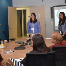
13th Annual Tony Cerenzia Research Lecture
Guest speaker Dr. Kidd’s talk – “From clinical trials to the clinic: A story about making Cognitive Adaptation Training for schizophrenia more accessible” – focused on how to implement interventions proven in clinical trials. Dr. Kidd is a clinical psychologist, senior scientist and psychology division chief at the Centre for Addiction and Mental Health (CAMH) in Toronto. He is also an associate professor in the Department of Psychiatry at the University of Toronto.
Image
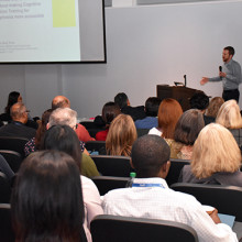
Above: Dr. Sean Kidd's lecture focused on implementing interventions proven in clinical trials.
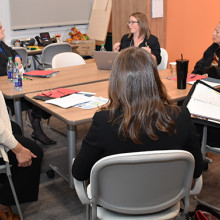
Workshop Sessions
Following Dr. Kidd’s lecture, attendees had the opportunity to participate in one of six workshops on a variety of topics related to recovery and rehabilitation focused mental health research:
“Implementing Interventions: A facilitated conversation attending to evidence, strategy, and recovery oriented care”
Led by Dr. Sean Kidd
Participants shared successful strategies for implementing novel approaches to care and discussed the challenges involved. They also looked at ways to leverage technology and education materials.
“Spirituality in Mental Health Care: Practically Supporting Recovery and Wellness”
Led by Stephen Yeo, Lawson allied scientist and chaplain, Southwest Centre; and Dr. Clark Heard and Jared Scott, Lawson associate scientists and occupational therapists, Southwest Centre
This workshop focused on the practical application of spirituality within the clinical setting, including the use of labyrinths, which contribute to recovery by promoting spiritual self-care, insight development and personal meaning-making reflection. Attendees had the opportunity to participate in a labyrinth walk and a related spiritual reflection. Read more about the labyrinths at Parkwood Institute and the Southwest Centre or watch the following video featuring highlights from the workshop:
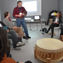
“Indigenous Men’s Health and Wellbeing: Connection with Culture as a Rehabilitation and Recovery Tool”
Led by Bill Hill, social worker, Parkwood Institute and Dr. Vicki Smye, associate professor, director of nursing at Western University
Through the sharing of practitioner experience and Indigenous men’s voices, the workshop focused on understanding the powerful links between connection to culture and mental health and well-being (pictured below).
Image
“Engaging Service Users and their Families in Research”
Led by Dr. Cheryl Forchuk, The Beryl and Richard Ivey Research Chair in Aging, Mental Health, Rehabilitation and Recovery, Lawson; and Deborrah Sherman, executive director, Ontario Peer Development Initiative
Participants in this workshop discussed the benefits of patient and family involvement in mental health research and identified strategies to support patient and family engagement (pictured below).
Image
“Innovation in Mental Health Care: Moving Ideas to Impact”
Led by Kaitlin Saxton, research and innovation facilitator, Parkwood Institute; and Lisa Bitcola, centre manager of projects and operations, Ivey International Centre for Health Innovation
This workshop focused on how innovation relates to research and quality improvement initiatives within St. Joseph’s Mental Health Care facilities. Participants discussed innovative approaches that could be implemented within their own clinical practice, research and quality improvement initiatives (pictured below).
Image
“My Professional Practice: Where's the Research?”
Led by Amanda Thibeault, director, professional practice, St. Joseph’s
In this session, participants discussed how they can incorporate research into their clinical practice (pictured below).
Image
Improving surgery for wrist arthritis
Wrist arthritis can cause debilitating pain, weakness and decreased range of motion. When patients are first diagnosed, the condition can often be managed with activity modification and pain medication. However, as symptoms progress, patients eventually require surgery.
Surgeons typically perform a procedure called four-corner fusion to preserve wrist motion and provide pain relief. This surgery involves removing one of the carpal bones and fusing four of the remaining carpal bones. Although this procedure is one of the most common treatments for wrist arthritis, it is not known how the position of the fusion of the wrist bones affects range of motion and joint contact.
Lawson associate scientist Dr. Nina Suh is leading a study with the goal of improving the surgical technique for four-corner fusion to maximize wrist function and symptom relief, and delay wrist arthritis progression.
Dr. Suh and her team will use a customized active-motion wrist simulator to create different carpal bone fusion positions. They will then assess how these positions affect wrist motion and joint contact area.
“We hope this research will lead to new surgical techniques that will help us to more effectively manage wrist arthritis with four-corner fusion,” says Dr. Suh, who is also an orthopaedic surgeon at the Roth McFarlane Hand and Upper Limb Centre (HULC) at St. Joseph’s Health Care London and an assistant professor at Western University’s Schulich School of Medicine & Dentistry. “The project will also advance our understanding of wrist biomechanics, providing a foundation for the development of enhanced patient-specific surgical tools, such as custom wrist fusion devices and implants.”
Image
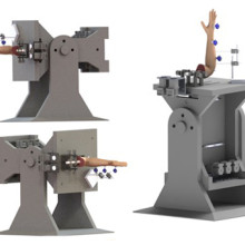
Image of the customized active-motion wrist simulator Dr. Nina Suh and her team are using to create different carpal bone fusion positions. They will then assess how these positions affect wrist motion and joint contact area.
The study is being funded through the Lawson Internal Research Fund (IRF), designed to allow scientists the opportunity to obtain start-up funds for new projects with exciting potential.
“The IRF program is valuable for scientists as external funding sources routinely require preliminary data to strengthen applications,” says Dr. Suh. “Particularly for new scientists such as myself, these grants provide seed funding that allows us to demonstrate the validity of our methodology and the clinical usefulness of our results.”
The IRF is designed to provide Lawson scientists the opportunity to obtain start-up funds for new projects with the potential to obtain larger funding, be published in a high-impact journal, or provide a clinical benefit to patients. Funding is provided by the clinical departments of London Health Sciences Centre and St. Joseph’s Health Care London, as well as the hospital foundations (London Health Sciences Foundation and St. Joseph’s Health Care Foundation).
Reviewed and Updated by: Jeff Forbes Emergency Management and Risk Specialist August 16, 2023 Incident Management System (IMS) Roles and Responsibilities Roles and Responsibilities Incident Manager o Administrator on Call or Delegate Role: o Overall authority and responsibility for the Emergency Ope...
… where incident may have taken place. Individuals in Bank: Patients or visitors who have or may have been injured. … 15 years after the date of the last discharge, or death. Patients … where incident may have taken place. Individuals in Bank: Patients or visitors who have or may have been injured. …
Personal Information Bank (PIB) Details Title: Incident Reports Location of Records: Risk Management Office, appropriate departmental files Description: Reports relating to incidents occurring in the hospital or on hospital property where a patient or visitor has been or may have been injured. Legal...
… justice system. Through intensive work with our care teams, patients are able to develop the new skills and supports … Health Care OUR PROGRAM OUR AIM EDUCATION AND TEACHING Patients within Southwest Centre’s Forensic Psychiatry … referred for assessment by the criminal justice system. All patients are under the jurisdiction of the Ontario Review …
CARING FOR THE BODY, MIND & SPIRIT SINCE 1869 Renowned for compassionate care, St. Josephs is one of the best academic health care organizations in Canada dedicated to helping people live to their fullest by minimizing the effects of injury, disease and disability through excellence in care, teachin...
… Complex Continuing Care: Information for Patients, Families, and Visitors Complex Continuing Care is … Care program serves three primary patient groups: Patients whose care needs exceed those that can be met in a … to meet the rehabilitation admission criteria; and, Patients with multiple care needs or disease processes who …
Complex Continuing Care: Information for Patients, Families, and Visitors Complex Continuing Care is a 62 bed inpatient service intended for adults whose ongoing complex care needs cannot currently be met in the community or in a long-term care facility. It is a part of the health care continuum des...
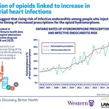
Injection of opioids linked to significant increase in bacterial heart infections
People who inject drugs are at a high risk for a number of health issues. In a new study from ICES, Lawson Health Research Institute and Western University, researchers discovered a significant rise in the risk of infective endocarditis, a serious heart infection, among Ontarians who inject drugs. When examining opioid prescriptions in the province, the research team discovered the increased risk of infective endocarditis may be related to the growing use of a specific opioid, hydromorphone.
The researchers looked at de-identified Ontario health data for 60,529 hospital admissions related to injection drug use between 2006 and 2015. Of the 60,529 admissions, 733 patients had infective endocarditis. Although hospital admission rates in people who inject drugs were stable over the study period, the risk of infective endocarditis increased from 13.4 admissions to 35.1 admissions every three months.
“Rates of infective endocarditis in people who inject drugs have been increasing around the world and our study shows this is true in Ontario,” says Dr. Matthew Weir, adjunct scientist at ICES, associate scientist at Lawson and assistant professor at Schulich School of Medicine & Dentistry, Western University. “We wondered if a change in the types of drugs people inject was responsible for this higher risk.”
Through further analysis of Ontario health data, the team discovered the increasing risk of infective endocarditis may be linked to a rise in prescriptions of the opioid hydromorphone. The number of hydromorphone prescriptions in Ontario increased from 16 per cent of all opioid prescriptions in 2006 to 53 per cent by 2015. This parallels the timing for increased risk of infective endocarditis among people who inject drugs.
Image
The researchers initially suspected the increased risk for infective endocarditis would begin when controlled-release oxycodone was removed from the market in 2011.
“We thought hydromorphone prescriptions would increase when controlled-release oxycodone was removed from the market, leading to increased risk of heart infection,” says Dr. Michael Silverman, associate scientist at Lawson and associate professor at Schulich Medicine & Dentistry. “However, while the study did show a substantial increase in risk for infective endocarditis, it began in 2010.”
Traditional controlled-release oxycodone was easily dissolvable and people who inject drugs did not save or reuse their injection equipment. Controlled-release hydromorphone, the more common form of the drug, is more difficult to dissolve. Since residue of the drug gets left in injection equipment, injection drug users save the equipment for future use or to share with others. Reusing injection equipment allows multiple opportunities for bacterial contamination, increasing the chances that bacteria will be injected when the equipment is next used.
Infective endocarditis occurs when the inner lining of the heart becomes infected. It can be a life-threatening illness and research suggests it can be caused by sharing or re-using injection equipment, possibly through the injection of bacteria.
“While the timing was not what we expected, we did find a correlation between the rise in infective endocarditis and hydromorphone prescriptions,” says Dr. Sharon Koivu, Lawson scientist and associate professor at Schulich Medicine & Dentistry. “Our research is now focused on better understanding the potential relationship between the injection of hydromorphone and risk of infective endocarditis.”
The team is conducting ongoing studies that are looking at whether bacteria that cause infective endocarditis are more likely to survive in equipment used to prepare hydromorphone compared to other drugs.
“The opioid crisis is one of the most pressing health issues of our time. Our findings not only confirm an increasing risk of infective endocarditis in persons who inject drugs but also offer the first evidence for why it might be happening,” says Dr. Weir. “Through research and collaboration, we hope to further collect the evidence needed to address this global problem.”
The study, “The risk of infective endocarditis among people who inject drugs: A retrospective, population-based time series analysis,” is published today in CMAJ (Canadian Medical Association Journal).
Scientist
Innovative ‘poop pills’ show promising results in clinical trials for multiple types of cancer
Two Canadian clinical trials show poop pills could help patients respond to immunotherapy while also reducing toxic side effects of cancer drugs
LONDON, ON and MONTREAL, QC – Fecal microbiota transplants (FMT), can dramatically improve cancer treatment, suggest two groundbreaking studies published in the prestigious Nature Medicine journal. The first study shows that the toxic side effects of drugs to treat kidney cancer could be eliminated with FMT. The second study suggests FMT is effective in improving the response to immunotherapy in patients with lung cancer and melanoma.
The findings represent a giant step forward in using FMT capsules – developed at Lawson Research Institute (Lawson) of St. Joseph’s Health Care London and used in clinical trials at London Health Sciences Centre Research Institute (LHSCRI) and Centre de recherche du Centre hospitalier de l’Université de Montréal (CRCHUM) – for safe and effective cancer treatment.
A Phase I clinical trial was conducted by scientists at LHSCRI and Lawson to determine if FMT is safe when combined with an immunotherapy drug to treat kidney cancer. The team found that customized FMT may help reduce toxic side effects from immunotherapy. The clinical trial involved 20 patients at the Verspeeten Family Cancer Centre at London Health Sciences Centre (LHSC).
“Standard treatment for advanced kidney cancer often includes an immunotherapy drug that helps the patient’s immune system tackle cancer cells,” says Saman Maleki, PhD, Scientist at LHSCRI. “But, unfortunately, the treatment frequently leads to colitis and diarrhea, sometimes so severe that a patient must stop life-sustaining treatment early. If we can reduce toxic side effects and help patients complete their treatment, that will be a gamechanger.”
Separate Phase II lung and skin cancer studies were led by researchers at CRCHUM in collaboration with Lawson and LHSCRI. The studies found that 80 per cent of patients with lung cancer responded to immunotherapy after FMT, compared to only 39-45 per cent typically benefiting from immunotherapy alone. Similarly, 75 per cent of patients with melanoma who received FMT experienced a positive response to treatment, compared to only 50-58 per cent response in patients who receive immunotherapy alone. Twenty patients participated in the lung cancer clinical trial and 20 patients participated in the skin cancer clinical trial.
“Our clinical trial demonstrated that fecal microbiota transplantation could improve the efficacy of immunotherapy in patients with lung cancer and melanoma,” says Dr. Arielle Elkrief, co-principal investigator and Physician Scientist, Université de Montréal-affiliated hospital research centre (CRCHUM). “The results also uncovered one possible mechanism of action of fecal transplantation—through the elimination of harmful bacteria following the transplant. Our results open up a novel avenue for personalized microbiome therapies, and fecal transplant is now being tested as part of the large pan-Canadian Canbiome2 randomized controlled trial.”
“Fecal microbiota transplantation in melanoma and lung cancer opens an entirely new therapeutic avenue, made possible by the exceptional commitment of our patients and the teamwork,” adds Dr. Rahima Jamal, Director of the Unit for Innovative Therapies (UIT) at CRCHUM. “At the Unit for Innovative Therapies (UIT) of the CRCHUM, we have had the privilege of translating laboratory discoveries into early phase clinical trials and witnessing their concrete impact on people living with cancer.”
Both studies use advanced, world-leading FMT capsules, also known as LND101, produced by Lawson in London, Ont. The research reinforces London’s place as a global leader in FMT innovation and treatment. The capsules are processed from healthy donor stools and ingested to help restore a patient’s healthy gut microbiome and treat different types of cancer.
“To use FMT to reduce drug toxicity and improve patients’ quality of life while possibly enhancing their clinical response to cancer treatment is tremendous, and it had never been done in treating kidney cancer before this,” says Dr. Michael Silverman, Scientist at Lawson and Head of St. Joseph’s Infectious Diseases Program. “And none of this would be possible if not for this close collaboration: innovating the FMT capsules in Lawson labs and introducing them at LHSCRI and CHUM to advance vital research initiatives. Also, because LND101 comes from healthy donors, production can be scaled up to eventually help large numbers of cancer patients.”
The studies build on earlier London and CHUM-generated Phase I research showing FMT can safely augment treatment for people with melanoma. FMT is also being studied in people with pancreatic cancer and triple-negative breast cancer, and is already a well-established treatment for serious gut infections such as C. difficile, which can cause severe diarrhea.
“Our hope is that our research will one day help people with cancer live longer while reducing the harmful side effects of treatment,” adds Dr. Ricardo Fernandes, Scientist at LHSCRI and Medical Oncologist at LHSC. “We are world leaders in FMT research and we’re excited about its potential.”
The lung and melanoma study was funded by the Canadian Cancer Society. The kidney cancer clinical trial was funded with support from Ontario Institute of Cancer Research, Canadian Institutes of Health Research, AMOSO, Western University’s Division of Medical Oncology, donors to London Health Sciences Foundation and St. Joseph’s Health Care Foundation, the Hesch Foundation and Weston Family Foundation.
-30-
A video with interviews with Maleki, Silverman and Fernandes is available here. Broll is also available.
To arrange interviews, contact:
Celine Zadorsky, Senior Media Relations Consultant, London Health Sciences Centre, (226) 927-2309, @email
Ellen Caracas, Media Relations, CHUM, (438)-346-0755, @email
About Université de Montréal-affiliated hospital Research Centre (CRCHUM). The CRCHUM is the Université de Montréal’s largest biomedical and health care science research centre and one of the most impressive and modern in all of Canada. Here, basic, clinical and population health research are carried out side-by-side under one roof. Find us online : www.chumontreal.qc.ca/en/crchum and on social media @CRCHUM
About Lawson Research Institute: Lawson Research Institute, the health innovation arm of St. Joseph's Health Care London, is committed to making and sharing discoveries that improve lives locally and internationally. Every day, Lawson researchers work to transform imagination to innovation to patient impact. Lawson leads health-care research. Find us online at sjhc.london.on.ca/research and on social media @stjosephslondon
About London Health Sciences Centre Research Institute: At London Health Sciences Centre Research Institute (LHSCRI), our teams pioneer discoveries that transform the health of adult and paediatric patients around the world. As the research institute of London Health Sciences Centre (LHSC), we conduct research where patient care is delivered, working alongside patients, families, health-care providers and academic partners like Western University. We are leaders in advancing the understanding, diagnosis, treatment and management of diseases and health conditions through a diverse research program that ranges from laboratory-based science to clinical trials. Our research has a global impact as we build on LHSC’s 150-year legacy of health innovation and drive forward medical breakthroughs that make a difference in the lives of patients and their families. Find us online at www.lhscri.ca and on social media @LHSCRI.