Search
Search
2373 Search Results:
… February 2019 Instructions for completing the health review form In order to … of your employment offer, the following information must be provided to Occupational Health and Safety Services no later than 7 business days prior to your start date. INCOMPLETE FORMS AND LATE SUBMISSIONS WILL DELAY …
February 2019 Instructions for completing the health review form In order to fulfill the terms and conditions of your employment offer, the following information must be provided to Occupational Health and Safety Services no later than 7 business days prior to your start date. INCOMPLETE FORMS AND L...
… October 2021 Instructions for completing the health review form Please … Safety Services no later than 7 business days prior to your start date. INCOMPLETE FORMS AND LATE SUBMISSIONS WILL DELAY YOUR START DATE. 1. Red Measles You require 2 doses of measles … October 2021 Instructions for completing the health review form Please …
October 2021 Instructions for completing the health review form Please provide the following information to Occupational Health and Safety Services no later than 7 business days prior to your start date. INCOMPLETE FORMS AND LATE SUBMISSIONS WILL DELAY YOUR START DATE. 1. Red Measles You require 2 d...
… Centre QUICK “RULE OF THUMB” FOR INSULIN ACTION INSULIN STARTS TO WORK IN: WORKS HARDEST AFTER: LASTS FOR: Fast-acting (bolus) Insulin Fiasp (insulin aspart) … Centre QUICK “RULE OF THUMB” FOR INSULIN ACTION INSULIN STARTS TO WORK IN: WORKS HARDEST AFTER: LASTS FOR: …
Diabetes Education Centre QUICK RULE OF THUMB FOR INSULIN ACTION INSULIN STARTS TO WORK IN: WORKS HARDEST AFTER: LASTS FOR: Fast-acting (bolus) Insulin Fiasp (insulin aspart) 5 minutes 45 60 minutes 3 5 hours Apidra (insulin glulisine) Humalog (insulin lispro) Humalog U200 (insulin lispro 200 uni...
Interdisciplinary team explores London’s unique approach to addressing homelessness
In London, Ont. more than 1,700 people are experiencing homelessness.
Recognizing the growing crisis, more than 200 individuals from 70 organizations across the city came together and developed the Health & Homelessness Whole of Community System Response – a strategic roadmap for supporting unhoused people who have the most complex health and social support challenges, and helping them access permanent housing that meets their needs. Now a team of researchers has been tasked with assessing how well the roadmap is working.
To understand if the program is meeting its ambitious goals and explore areas for improvement, the Centre for Research on Health Equity and Social Inclusion (CRHESI), a community-university partnership based at Western’s Faculty of Health Sciences, was asked by the Whole of Community Response leaders to support and coordinate research and evaluation of the program.
CRHESI works with and reports to the System Foundation Table, a team of volunteer community partners from diverse sectors with a mandate to evaluate new activities and embed continuous improvement to ensure sustainability of the program.
“London’s Whole of Community Response is a novel approach to addressing the needs of the most vulnerable individuals experiencing complex housing and health challenges within our community,” said Mick Kunze, chair of the System Foundation Table. “While our approaches and interventions are rooted in evidence-based practices, we also understand that local context matters.”
This research and evaluation work is funded jointly by Western, London Health Sciences Centre and through a generous donation from local businessman Ryan Finch to St. Joseph’s Health Care Foundation. The three groups came together recognizing there is a need to create meaningful and measurable change in our community.
Their support has allowed CRHESI to recruit two research managers, Eleanor Gebrou and Kelly Barnes, who oversee the work of more than 100 researchers and community partners from organizations across the city participating in the evaluation project.
"Community-led initiatives not only reflect the real needs of individuals facing homelessness, but they also empower frontline workers by fostering an environment of collaboration and understanding. Together, we can create solutions that prioritize well-being for all—those we serve and those who serve," said Gebrou.
This work is split into four main research areas:
- Exploring the outcomes and experiences of people who are precariously housed, unhoused or at risk of homelessness, as well as the perspectives of the broader residents of London, Ont. and especially the business community.
- Evaluating the experiences and well-being of those working in jobs that provide care and housing support.
- Answering questions related to systems, structures, processes and costs of care.
- Evaluating the processes that enabled this large and complex “whole of community” response, and how it unfolds from here.
“I really believe in evidence-based decision making and having as much information as we can to go into program and policy design,” said Barnes. “It's so important to understand what parts of our homelessness response are working well and what can be improved. Research and evaluation are some of the best ways to do that.”
The importance of voices and stories
Research teams will collect quantitative data, including statistics on how many people get housed, how this housing affects the number of hospital and police contacts and the cost-benefit of these new activities. They’ll also collect qualitative data, which involves hearing the experiences of those without homes, those accessing care and support and those providing and leading services.
This evaluation framework will be presented to city council, sitting as the Strategic Priorities and Policies Committee, on October 8, 2024.
“It is really important to us that this not just be numbers,” said Barnes. “We know how important people’s voices and stories are. When we take the numbers and combine them with people’s descriptions of their experiences, and the discussion of their journey, that gives us really solid information to bring back to the policy-makers and program developers to show what’s working and what isn’t.”
This research and evaluation work will span the next two years, with results shared annually with city council starting in July 2025. In addition to annual reporting, research project teams will share data as it emerges.
“The research and evaluation efforts led by the backbone team of this movement, including the System Foundations Table and our CHRESI research and evaluation managers, will be instrumental in guiding our present and future work, by illustrating the real impact our interventions are having on the health and well-being of those we’re aiming to support, as well as by responding to the questions and concerns voiced by all members of our community,” said Kunze.
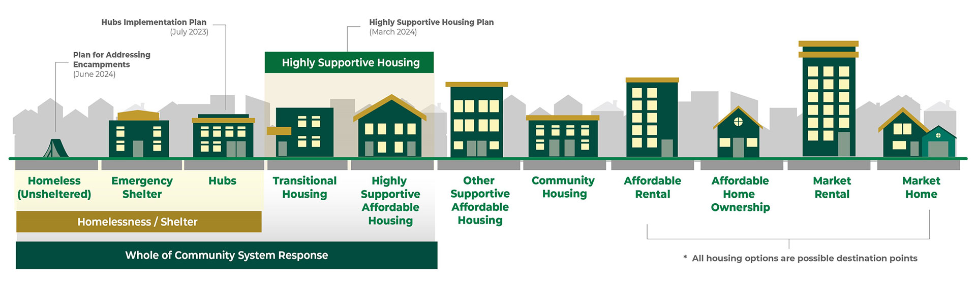
Health & Homelessness Whole of Community System Response
The Whole of Community Response is about developing wrap-around health care and housing supports for those who need them most, ensuring basic human needs are met and building trusting relationships with people so they’re set up to succeed. It includes three main pillars: establishing hubs to meet immediate needs for safe shelter, nutrition and hygiene, and allowing care providers to start the process of stabilizing people’s mental and physical health. Alongside establishing hubs, the broader plan includes bringing more highly supportive housing units to London and implementing a human rights-based approach to support people wherever they are on the housing spectrum.
City council endorsed the Whole of Community Response approach in March 2023. Since then, two hubs have been established, as well as 93 highly supportive housing units, with 50 more units in development, toward a goal of 600 highly supportive housing units within three years.
The Whole of Community Response is being facilitated through the City of London and implemented by lead agencies, with ongoing collaboration among several sectors, including police and emergency services, hospitals, front-line community service organizations, educational institutions and government.
The Health and Homelessness Fund for Change, fuelled by a transformative $25-million donation from a London, Ont. family who wishes to remain anonymous, primarily provides capital funding to help fast-track the creation of hubs and highly supportive housing units. The Fund for Change is administered by London Community Foundation in partnership with the donor family.
… physician(s) you feel should assess patient, Otherwise first available Lillian Barra MD, FRCPC □ Rheumatologist William F. Clark, MD, FRCPC □ Nephrologist Michael Strong, MD, FRCPC □ Neurologist INTERDISCIPLINARY … physician(s) you feel should assess patient, Otherwise first available Lillian Barra MD, FRCPC □ Rheumatologist …
Select physician(s) you feel should assess patient, Otherwise first available Lillian Barra MD, FRCPC Rheumatologist William F. Clark, MD, FRCPC Nephrologist Michael Strong, MD, FRCPC Neurologist INTERDISCIPLINARY VASCULITIS CLINIC REFERRAL FORM Fax referral form to: 519-646-6072 Patient Informat...
… St. Joseph’s Hospital Diagnostic Imaging Centre, Room C0-200 268 Grosvenor St., PO Box 5777, Stn. B London, ON N6A 4V2 Tel. 519-646-6044 INTERVENTIONAL … St. Joseph’s Hospital Diagnostic Imaging Centre, Room C0-200 268 Grosvenor St., PO Box … Interventional radiology request form …
St. Josephs Hospital Diagnostic Imaging Centre, Room C0-200 268 Grosvenor St., PO Box 5777, Stn. B London, ON N6A 4V2 Tel. 519-646-6044 INTERVENTIONAL RADIOLOGY REFERRAL FORM Fax to 519-646-6204 1. Patient Information Last name: ______________________ First Name: ____________________ Date of birth: ...
… Hodges TORONTO | Critics of the Canadian Association of Gastroenterology’s (CAG) recently updated position state- ment on colorectal cancer screening are slamming the … “To call this colorectal cancer screen- ing is dishonest,” Dr. Patricia Johans- son, a family physician in …
by David Hodges TORONTO | Critics of the Canadian Association of Gastroenterologys (CAG) recently updated position state- ment on colorectal cancer screening are slamming the new recommenda- tion that flexible sigmoidoscopyand not colonoscopybe offered to all average-risk individuals. To call this c...
… FOR INTRAVENOUS FLUORESCEIN ANGIOGRAPHY You have been requested to have the Ophthalmic Diagnostic test INTRAVENOUS FLUORESCEIN ANGIOGRAPHY performed in the St. … FOR INTRAVENOUS FLUORESCEIN ANGIOGRAPHY You have been requested to have the Ophthalmic Diagnostic test INTRAVENOUS …
PATIENT CONSENT FOR INTRAVENOUS FLUORESCEIN ANGIOGRAPHY You have been requested to have the Ophthalmic Diagnostic test INTRAVENOUS FLUORESCEIN ANGIOGRAPHY performed in the St. Josephs Hospital What is uorescein angiography ? Fluorescein angiography is a diagnostic procedure which uses a special came...
Introducing The Gray Centre for Mobility and Activity at Parkwood Institute
12:00 PM - 01:00 PM
Join us Thursday October 21 from 12 pm – 1 pm for a virtual introduction to some of the exciting research and clinical innovations from mobility experts at The Gray Centre for Mobility and Activity. Hear presentations from:
- Siobhan Schabrun, PhD – Harnessing the Brain to Reduce Pain and Improve Mobility and Activity.
- Sue Peters, PhD – Wireless Neuroimaging During Mobility to Predict Recovery Trajectories after Stroke.
- Swati Mehta, PhD – Virtual Physical Activity Programming During the Pandemic.
- Dr. Manuel Montero-Odasso – Mobility and Cognition. The Collision of 2 Giants.
- Stephanie Cornell, MPT – Who, What, When? Using Technology in Rehab.
About The Gray Centre for Mobility and Activity:
Established in 2020, The Gray Centre for Mobility and Activity at St. Joseph’s Health Care London is expanding and advancing mobility and rehabilitation treatment and prevention solutions through research, collaborations and the latest technology to improve the lives of those living with disease, disability or injury. The Gray Centre is located at St. Joseph’s Parkwood Institute, Southwestern Ontario’s regional provider of rehabilitation and recovery health care and a national hub for treatment, research and education in mobility and activity. The Gray Centre is made possible through a $7.5 million gift to St. Joseph’s Health Care Foundation from William and Lynne Gray.
Partners
Introducing The Gray Centre for Mobility and Activity at Parkwood Institute
4:00pm - 5:00pm
Image

Image
Join us Thursday October 21 from 12 pm – 1 pm for a virtual introduction to some of the exciting research and clinical innovations from mobility experts at The Gray Centre for Mobility and Activity. Hear presentations from:
- Siobhan Schabrun, PhD – Harnessing the Brain to Reduce Pain and Improve Mobility and Activity.
- Sue Peters, PhD – Wireless Neuroimaging During Mobility to Predict Recovery Trajectories after Stroke.
- Swati Mehta, PhD – Virtual Physical Activity Programming During the Pandemic.
- Dr. Manuel Montero-Odasso – Mobility and Cognition. The Collision of 2 Giants.
- Stephanie Cornell, MPT – Who, What, When? Using Technology in Rehab.
About The Gray Centre for Mobility and Activity:
Established in 2020, The Gray Centre for Mobility and Activity at St. Joseph’s Health Care London is expanding and advancing mobility and rehabilitation treatment and prevention solutions through research, collaborations and the latest technology to improve the lives of those living with disease, disability or injury. The Gray Centre is located at St. Joseph’s Parkwood Institute, Southwestern Ontario’s regional provider of rehabilitation and recovery health care and a national hub for treatment, research and education in mobility and activity. The Gray Centre is made possible through a $7.5 million gift to St. Joseph’s Health Care Foundation from William and Lynne Gray.
Investing in life-changing research
Through donor support, endowed research chairs are exploring and answering some of the most profound and complex research questions of our time.
Among cherished family photos and special mementos in the office of Jeremy Burton, PhD, is a slightly faded photo of a young woman. Burton points out the framed photo as he enthusiastically talks about his work. It’s a young Miriam Burnett, after whom the Miriam Burnett Chair in Urological Sciences is named. It’s also the first endowed research chair position Burton held at St. Joseph’s Health Care London (St. Joseph’s).
As the research chair for seven years, Burton speaks fondly about the relationship he has with the Burnett family and the crucial role their support has played in advancing his research.
“Thanks to their funding, we became one of the world leaders in urological microbiome research,” he says.
Endowed research chairs at St. Joseph’s receive consistent and sustainable funding so that research leaders and their teams can answer the most profound and complex health questions of our time.
For decades, donors have been inspired by the clinical research taking place at St. Joseph’s and have heavily invested in endowed research chairs. Today, St. Joseph’s Health Care Foundation manages seven endowed chairs focused on several areas, including molecular imaging, fetal and newborn growth and diabetes. Working in partnership with Western University, and with donor support, the foundation recently established four new endowed chairs in mobility, medical biophysics, medical imaging and ophthalmology.
“Medical research in Canada is chronically underfunded, and there is almost no sustainable funding for hospital-based research positions,” says Michelle Campbell, President & CEO, St. Joseph’s Health Care Foundation. “Private philanthropy has filled that gap for years. When a donor gives to an endowed research chair, they are building capacity in the present day and creating future value and opportunity. An endowed gift has a multiplier effect.”
Burton, now the endowed Research Chair in Human Microbiome and Probiotics, has many reasons to be grateful for this support. Not only does the endowed fund pay for Burton’s research salary, it also partially supports the salaries of a lab manager and technical team – all vital for a sophisticated lab to be successful.
The funding also provides the gift of time – a diminishing commodity for any busy research team.
“Scientists need more time to think,” says Burton, a Lawson Research Institute (Lawson) scientist. “We are incrementally being stretched in multiple directions, and the funding gives us the time to do what we are meant to do – find answers to important clinical questions and find solutions to medical problems.”
Distinguished Lawson scientist and university professor Cheryl Forchuk, PhD, wholeheartedly agrees. She recently completed her final term as The Beryl and Richard Ivey Research Chair in Aging, Mental Health, Rehabilitation & Recovery, another endowed position. As Chair, Forchuk provided scientific and administrative leadership to a large group of researchers based at St. Joseph’s Parkwood Institute focused on mental health, activity and mobility, and cognitive vitality and brain health.
Many research leaders, she explains, can afford to spend only two days a week on their own research projects. Endowed chair positions change that.
“Imagine travelling across the country to create a national study focused on homelessness, two days a week at a time,” she suggests candidly. “You couldn’t.”
Forchuk is referring to her landmark project to better understand how many people in Canada are homeless and who they are. The goal was to develop more accurate sources of data and recommend appropriate support and services. Her work is already resulting in important changes.
Today, Forchuk is embarking on another cross-country research project to find solutions related to homelessness for Canadian veterans who are women.
Like Forchuk, Burton’s Chair position requires him to provide operational and research leadership, including developing research networks and partnerships nationally and internationally to advance studies that will revolutionize care.
“As the Chair, I think it is important that I have wide-ranging projects that benefit people in our own community and beyond,” says Burton, who is optimistic about the outcomes of several of his team’s studies.
He recently partnered with London’s First Episode Mood and Anxiety Program to study the impact of fermented foods on the microbiome of young people taking medications for mental health conditions.
One of the side effects of these medications is weight gain, which deters some patients from taking it. By providing patients with slow-release apple cider capsules, which have similar properties to fermented foods and positively affect the microbiome, they have seen an overall improvement in participants’ mental health and cholesterol after just a few months.
Reflecting on his team’s research achievements to date and the potential of what’s to come, Burton emphasizes how vital endowed chairs are to the sustainability of research and the hope to translate newly discovered knowledge into medical practice.
“Research funding from other sources comes and goes,” he says, “but endowed chair positions that are focused on improving human health provide continuity, build research and create change benefiting all of us.”
iSee Vision Screening Program - Screening Session and Special Gift Announcement
1:30pm
Join the St. Joseph's Health Care Foundation and the Brandon Prust Foundation for a special gift announcement at the Stoney Creek YMCA on Monday, July 18 at 9:30 AM. This is also an opportunity to screen your children (aged 18 months to 4 years) for early signs of vision impairment through the iSee Vision Screening Program. Learn about the iSee Program by visiting the St. Joseph's Health Care London website.
… Friday, April 23, 2010 First Floor ‐ 300 York Street London, Ontario N6B 1P8 … • To update knowledge on new diagnostic and new therapeutic techniques • … Friday, April 23, 2010 First Floor ‐ 300 York Street …
CLINICALDAYINOPHTHALMOLOGY2010 OPHTHALMICPROBLEMSFRONTANDBACK LondonConventionCentreSalonC,D,E Friday,April23,2010 FirstFloor300YorkStreet London,OntarioN6B1P8 GenerouslySupportedBy: TheCharlesDysonLectureshipinOphthalmology TheRoyalCollegeofPhysicians&SurgeonsofCanada The...
… St. Joseph’s Health Care London ORTHOPTIST - IVEY EYE INSTITUTE 268 Grosvenor Street, Room B1-409 London, Ontario, … St. Joseph’s Health Care London ORTHOPTIST - IVEY EYE INSTITUTE 268 Grosvenor Street, Room B1-409 …
St. Josephs Health Care London ORTHOPTIST - IVEY EYE INSTITUTE 268 Grosvenor Street, Room B1-409 London, Ontario, Canada N6A 4V2 Telephone: 519 646-6018 Fax: 519 646-6056 REQUEST FOR ORTHOPTIC ASSESSMENT PATIENT: _________________________________________________________________________ DATE OF APPO...
… CME Sessions A Follow-up Evaluation at the University of Western Ontario Laeeq Tahir, MD, FRCP(C)1 , Nicole Tsang, MD2, … professional development ____________________ 1 Assistant Professor, Clinical Psychiatry, Dalhousie University and Consultant Psychiatrist, The Moncton Hospital, Moncton, New Brunswick. At the …
Participant Learning at Psychiatry CME Sessions A Follow-up Evaluation at the University of Western Ontario Laeeq Tahir, MD, FRCP(C)1 , Nicole Tsang, MD2, Marnin Jori Heisel, PhD, CPsych3 *, Jatinder Takhar, MD, FRCP(C)4 Key Words: continuing medical education, CME, knowledge transfer, professional ...
… Sciences Centre (hereafter referred to as “LHSC”) and St. Joseph’s Health Care, London (hereafter referred to as “St. Joseph’s”) IN CONSIDERATION of the mutual covenants and … covenant and agree to jointly govern and manage all existing and future voluntary arrangements and relationships …
Joint Collaboration Agreement between London Health Sciences Centre (hereafter referred to as LHSC) and St. Josephs Health Care, London (hereafter referred to as St. Josephs) IN CONSIDERATION of the mutual covenants and other good and valuable consideration contained herein, the parties covenant and...
… Boards of Directors of London Health Sciences Centre and St. Joseph’s Health Care, London approved the following … to facilitate the implementation of the Health Services Restructuring commission’s directives for London. In 1999, the … and sharing between London Health Sciences Center and St. Joseph’s Health Care, London. 1. We will drive decisions …
ICL Doc #20825 Principles for the Relationship of Londons Hospitals In 1997, the Boards of Directors of London Health Sciences Centre and St. Josephs Health Care, London approved the following principles. Their purpose was to facilitate the implementation of the Health Services Restructuring commiss...
… Dr. will provide 50% of the secretarial computer cost. You will be expected to lease/purchase the remaining … “A Guide to Secretarial Support Services for Professional Staff” at https://intra.lhsc.on.ca/medical-affairs/professional-staff/resources/secretarial-support for more information. …
Dr. s copy Western Schulich School of Medicine & Dentistrys copy Department of s copy Medical Affairs copy THIS LETTER OF OFFER IS ONLY TO BE ISSUED AFTER CONFIRMATION OF THREE SATISFACTORY REFERENCES FOR THE CANDIDATE. Dear Dr.
Dr. s copy Western Schulich School of Medicine & Dentistrys copy Department of s copy Medical Affairs copy THIS LETTER IS ONLY TO BE ISSUED TO CONFIRM A CHANGE IN PRIVILEGES OR ARC CATEGORY TO AN EXISTING CREDENTIALED CLINICAL ACADEMIC