Illuminating discovery 126 years ago shapes medical care today
The first-ever Nobel Prize in Physics was awarded to Wilhelm Röntgen in 1901 in recognition of the extraordinary services he rendered by the discovery of the “remarkable rays” subsequently named after him."
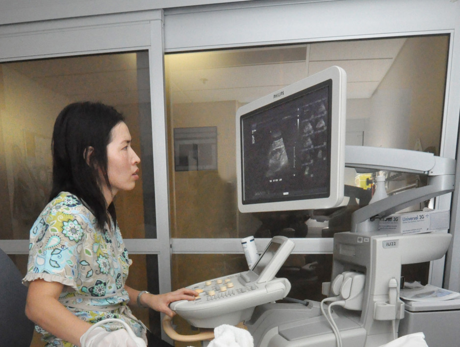
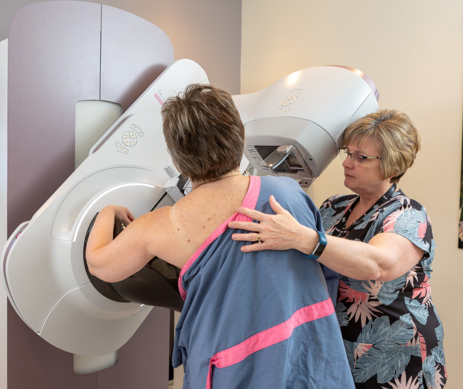
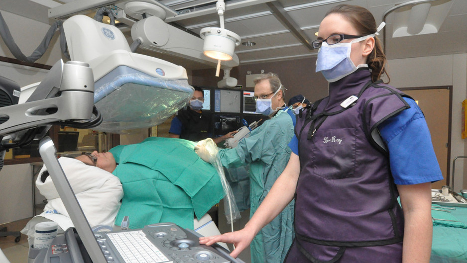
Whether you call them “remarkable rays”, “Röntgen rays” or “X-rays”, it is undeniable that Röntgen’s discovery on Nov. 8, 1895, has changed how we look at, and into, medical care. Patients at St. Joseph’s benefit from this discovery daily, with 93 registered medical radiation technologists (MRTs) performing more than 155,326 exams per year across multiple imaging modalities including X-rays, CT, nuclear medicine, MRI, PET/CT, mammography, interventional radiology, bone mineral density and ultrasound.
MRTs use their expert knowledge of imaging and/or radiation therapy equipment, together with an extensive understanding of the principles of anatomy, physiology and pathology, image acquisition, treatment and radiation protection to deliver quality care to patients, contributing their expertise to the diagnosis and treatment of millions of Canadians each year.
To mark Medical Radiation Technologist Week this year, MRTs at St. Joseph’s have collected some “illuminating” fun facts about the profession of medical imaging to shed light on the important role MRTs play in the care of patients:
Did you know…
- More common than you know: X-rays are more common throughout our life than you may know. Each year we are exposed to small amounts of naturally occurring radiation all around us. Some naturally occurring radiation include terrestrial radiation created by natural breakdown or decay of materials within earth, such as radon. Another form of naturally occurring radiation is cosmetic radiation, which are solar and galactic radiation from the sun/stars. We even receive small amounts of natural radiation from radionucleoids in everyday foods such as bananas, nuts and carrots! As well, everyday activities such as air travel and tanning contribute to our annual radiation dose.
- Technician vs technologist: There is a difference between an X-ray technician and X-ray technologist. An X-ray technician specializes in repairing/maintaining X-ray machines in addition to running calibration tests to ensure that medical imaging machines meet the province’s strict safety standards for use on patients. An X-ray technologist is a health care professional who has direct contract with patients and works to produce diagnostic images for the benefit of patients’ health.
- It's a photon - not image: Contrary to popular belief, the word X-ray refers to the form of photon energy used to obtain diagnostic images and not necessarily to the image itself. A radiograph is the resultant image that health care professionals and patients see after a patient has been exposed to X-rays. If you refer to the images as radiographs will be sure to impress those around you during your next visit!
- X-rays everywhere: After the discovery of X-rays in 1895, they were commonly used for everyday day activities up until the mid 1900s. For example, it was common to see X-ray machines in shoe stores. Customers, including young children, would try on shoes and put their feet into a X-ray machine to see how well the shoes fit. It was also common to see X-ray machines at carnivals where people could put their extremities into the machine to see their bones. At one time, people even used X-rays as a method to remove unwanted hair. This was all before the risks were known about prolonged exposure to radiation.
- At the speed of light: X-rays cannot be seen by the naked eye and cannot be felt. As the exposure is taken, X-ray photons travel at the speed of light and pass through the body. Energy from the X-rays are absorbed at different rates by different parts of the body to produce an image.
- Most important invention: In 2009, the discovery of X-ray was voted the most important scientific invention in a poll marking the centenary of the Science Museum in London, England, Britain, beating out penicillin, the DNA double helix structure and other inventions from past centuries in science, technology and engineering. Nearly 50,000 people voted.
- What's with the 'x' in X-ray: When German physicist Wilhelm Roentgen discovered a new form of radiation in 1895, he called it X-radiation because he didn't know what it was. He temporarily called them “X-rays,” with the “x” being a mathematical symbol for something unknown. To this day, the name stuck, though X-rays are occasionally called Roentgen rays in some countries.
Learn about St Joseph’s Medical Imaging Program
Medical imaging at St. Joseph's Health Care London supports and provides patient care using equipment and techniques that are at the technological forefront of medical imaging and intervention. Learn about the programs and services offered by the Medical Imaging Program.


