Search
Search
1376 Search Results:
… Dr. … Dr. …
Dr. s copy Western Schulich School of Medicine & Dentistrys copy Department of s copy Medical Affairs copy THIS LETTER IS ONLY TO BE ISSUED TO NON CLINICAL ACADEMIC APPOINTMENTS SUCH AS TERM OR LOCUM CATEGORY. Dear Dr.
… Dr. … Dr. …
Dr. s copy ( Western Schulich School of Medicine & Dentistrys copy ( Department of s copy ( Medical Affairs copy ( THIS LETTER IS ONLY TO BE ISSUED TO NON CLINICAL ACADEMIC APPOINTMENTS SUCH AS TERM OR LOCUM CATEGORY. Dear Dr.
… Dr. … Dr. …
Dr. s copy ( Western Schulich School of Medicine & Dentistrys copy ( Department of s copy ( Medical Affairs copy ( THIS LETTER IS ONLY TO BE ISSUED TO NON CLINICAL ACADEMIC APPOINTMENTS SUCH AS TERM OR LOCUM CATEGORY. Dear Dr.
… Dr. … Dr. …
Dr. s copy Western Schulich School of Medicine & Dentistrys copy Department of s copy Medical Affairs copy THIS LETTER IS ONLY TO BE ISSUED TO NON CLINICAL ACADEMIC APPOINTMENTS SUCH AS TERM OR LOCUM CATEGORY. Dear Dr.
… Dr. … Dr. …
Dr. s copy Western Schulich School of Medicine & Dentistrys copy Department of s copy Medical Affairs copy THIS LETTER IS ONLY TO BE ISSUED TO NON CLINICAL ACADEMIC APPOINTMENTS SUCH AS TERM OR LOCUM CATEGORY. Dear Dr.
… Dr. will provide 50% of the secretarial computer cost. You … as indicated for your files, and return all other copies to Dr. ’s office in the enclosed envelope. We would ask … Dr. will provide 50% of the secretarial computer cost. You … as indicated for your files, and return all other copies to Dr. ’s office in the enclosed envelope. We would ask …
Dr. s copy Western Schulich School of Medicine & Dentistrys copy Department of s copy Medical Affairs copy THIS LETTER OF OFFER IS ONLY TO BE ISSUED AFTER CONFIRMATION OF THREE SATISFACTORY REFERENCES FOR THE CANDIDATE. Dear Dr.
… Dr. Candidate’s copy Western Schulich School of Medicine & … of Oncology’s copy Medical Affairs’ copy DATE Dear Dr. Re: Letter of Understanding It gives us great pleasure … retirement. Letters of resignation/retirement should be addressed and sent to the Department of Chair/Chief with a …
Dr. Candidates copy Western Schulich School of Medicine & Dentistrys copy Department of Oncologys copy Medical Affairs copy DATE Dear Dr. Re: Letter of Understanding It gives us great pleasure to offer you a position as a General Practitioner in Oncology (GPO) in the Depart...
… Dr. … Dr. …
Dr. s copy Western Schulich School of Medicine & Dentistrys copy Department of s copy Medical Affairs copy THIS LETTER IS ONLY TO BE ISSUED TO CONFIRM A CHANGE IN PRIVILEGES OR ARC CATEGORY TO AN EXISTING CREDENTIALED CLINICAL ACADEMIC
… Dr. … Dr. …
Dr. s copy Western Schulich School of Medicine & Dentistrys copy Department of s copy Medical Affairs copy THIS LETTER IS ONLY TO BE ISSUED TO NON CLINICAL ACADEMIC APPOINTMENTS SUCH AS TERM OR LOCUM CATEGORY. Dear Dr.
… Dr. will provide 50% of the secretarial computer cost. You … as indicated for your files, and return all other copies to Dr. ’s office in the enclosed envelope. We would ask … Dr. will provide 50% of the secretarial computer cost. You … as indicated for your files, and return all other copies to Dr. ’s office in the enclosed envelope. We would ask …
Dr. s copy ( Western Schulich School of Medicine & Dentistrys copy ( Department of s copy ( Medical Affairs copy ( THIS LETTER OF OFFER IS ONLY TO BE ISSUED AFTER CONFIRMATION OF THREE SATISFACTORY REFERENCES FOR THE CANDIDATE. Dea...
… Dr. will provide 50% of the secretarial computer cost. You … as indicated for your files, and return all other copies to Dr. ’s office in the enclosed envelope. We would ask … issued by Western University. Chair/Chief, Department of Dr. William J. Sischek MD, FRCPC, CCPE Department of …
Dr. s copy ( Western Schulich School of Medicine & Dentistrys copy ( Department of s copy ( Medical Affairs copy ( THIS LETTER OF OFFER IS ONLY TO BE ISSUED AFTER CONFIRMATION OF THREE SATISFACTORY REFERENCES FOR THE CANDIDATE. Dea...
… Dr. … Dr. …
Dr. s copy ( Western Schulich School of Medicine & Dentistrys copy ( Department of s copy ( Medical Affairs copy ( THIS LETTER IS ONLY TO BE ISSUED TO NON CLINICAL ACADEMIC APPOINTMENTS SUCH AS TERM OR LOCUM CATEGORY. Dear Dr.
… Dr. … Dr. …
Dr. s copy ( Western Schulich School of Medicine & Dentistrys copy ( Department of s copy ( Medical Affairs copy ( THIS LETTER IS ONLY TO BE ISSUED TO NON CLINICAL ACADEMIC APPOINTMENTS SUCH AS TERM OR LOCUM CATEGORY. Dear Dr.
… Mental Health Care London, London, ON Corresponding author: Dr. Verinder Sharma, Specialized Adult Program, Regional … disorder. In spite of trials of various psychotropic drugs and frequent, prolonged hospitalizations, the patient … disorder and an emerging bipolar disorder 6. Subsyndromal symptoms that occur interepisodically in patients …
_______________________________________________________________________________________________________________ Case Report CONSEQUENCES OF THE MISDIAGNOSIS OF BIPOLAR DISORDER AS BORDERLINE PERSONALITY DISORDER by Hyacinth John, MD1, Verinder Sharma, MB, BS, FRCP(C) 2, 3 1 Resident, Department of P...
… forward from past meetings / business the board has addressed before) 4. New Business (20 mins) 4.1 Governance … Recognition of 2022/23 Retiring Board Members (5 mins) • Dr. Brian Rotenberg • Margaret Kellow 5. In-Camera Meeting … forward from past meetings / business the board has addressed before) 4. New Business (20 mins) 4.1 Governance …
Regrets to Terri-Lynn Cook 519-646-6100 ext. 64202 or terrilynn.cook@sjhc.london.on.ca P a g e | 1 Meeting of the Board of Directors Wednesday June 21, 2023 12:30 pm start time Parkwood Institute Main Bldg, Room A2-109A/B AGENDA REMINDER: At the forefront of our discussions today, we are to ensure t...
Keeping an eye on care of the future
Dr. Khaldon Abbas is using his curiosity and passion for ophthalmology to improve patient care and outcomes for people with eye diseases and disorders.
While in university, Dr. Khaldon Abbas had a deeply moving experience as a volunteer with the Canadian Centre for Victims of Torture (CCVT) that changed the trajectory of his life and career.
The community-based organization helps victims of war and torture, and Abbas, whose family immigrated to Canada from Iraq a little more than a decade before, wanted to share his skills as a translator and tutor with newcomers.
“I came to Canada when I was 12. I had limited English, we had no family or friends here, and it was really hard to acclimate,” says Abbas. “I wanted to give back to the community and to be there for immigrant families who were facing similar challenges that my family had to deal with.”
During one shift with CCVT, Abbas was paired with a family from Syria, whose nine-year old daughter was losing her eyesight. She was living with retinal dystrophy, a degenerative disorder that can progress to complete blindness.
Witnessing the impact the eye disorder had on the young girl and her family inspired Abbas to further his own education and set a goal to become an ophthalmologist.
That was eight years ago. Since then, Abbas spent several years working as a clinical research coordinator and completed four years of medical school at the University of British Columbia.
Today, he is a clinical research fellow at the Ivey Eye Institute of St. Joseph’s Health Care London (St. Joseph’s) – a position supported through St. Joseph’s Health Care Foundation thanks to the generosity of donors.
During the next year, Abbas’ research will focus on improving patient care and outcomes for people with eye diseases and disorders.
Drs. Phil Hooper, Verena Juncal and Tom Sheidow, all retinal surgeons at Ivey Eye, are the impetus behind the fellowship and now serve as Abbas’ mentors. Through the fellowship, the trio wanted to expand their clinical research program which is heavily focused on clinical trials. Their goal was to delve into quality improvement projects and explore, among other things, patient data, referral patterns and wait times – information that could guide Ivey Eye in refining care to better understand how to improve the overall flow of patient care.
As surgeons at the largest single-site eye care centre in Canada committed to innovative care, the Ivey Eye physician leaders felt a responsibility to make this work a reality.
“We started talking about this about three years ago,” says Sheidow. “We were familiar with similar roles at other academic eye care centres and we were fortunate to have some funding, so we brought the idea to the foundation and started to craft the terms of reference,” he adds.
Abbas is the second physician in this fellowship, following in the footsteps of Dr. Amy Basilious, who is now in her second year of residency at Ivey Eye.
“Amy did an exceptional job as our inaugural fellow and we were looking for someone with similar characteristics – bright, curious, motivated, a self-starter and a passion for ophthalmology,” says Sheidow. “Khaldon has all of that and more,” he adds, referring to Abbas’ interest and background in clinical trial work.
Even before arriving in London for the fellowship, Abbas began working with his new team to generate research project ideas and shape a research plan. Among the projects he will tackle is one that will assess the effectiveness and complications of lens exchange surgeries, and another in collaboration with Basilious focused on macular hole repairs.
He will also spearhead two quality improvement studies aimed at streamlining the referral process to Ivey Eye for optometrists and enhancing education and information resources for patients with eye diseases and disorders. Through his work, Abbas is excited to build his research skills, forge new professional connections and see some of his research translated into tangible improvements in patient care.
He’s grateful to Hooper, Juncal and Sheidow, along with St. Joseph’s and the Foundation, for their vision and spirit of innovation in establishing the fellowship.
“Everyone has been extremely welcoming and supportive of me, especially my mentors and fellow co-workers” he says. “There’s a real family environment at St. Joseph’s. I feel like this is my new home away from home.”
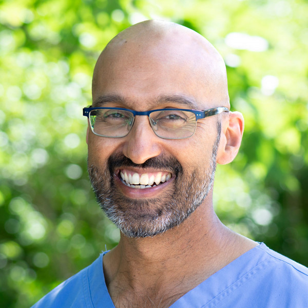
Keith Sequeira
Keith Sequeira, MD
Associate Professor, Schulich School of Medicine and Dentistry
Key Areas of Focus:
Implementation Science and Education
Phone:
519-685-4292 ext. 44029
Email:
Publications:
Accepting Grad Students:
No
About:
Dr. Keith Sequeira is a physiatrist at St. Joseph's Health Care London and London Health Sciences Centre and an associate professor in the Department of Physical Medicine and Rehabilitation in the Schulich School of Medicine and Dentistry at Western University. Dr. Sequeira completed his medical degree at the University of Toronto in 1994, residency training in Physical Medicine and Rehabilitation at Albany Medical Center in 1998, followed by a fellowship in Electrodiagnostic and Sports Medicine at Michigan State University.
Dr. Sequeira is the Medical Director of the Acquired Brain Injury Rehabilitation Program at Parkwood Institute and runs spasticity, EMG, brain and spinal cord injury clinics. Dr. Sequeira is the past Residency Program Director of Physical Medicine & Rehabilitation at Western, a program that he designed and initiated in 2005 and functions. He is the WSIB Champion, working on the integration of WSIB education into the medical school curriculum at Western, past director of the undergraduate the musculoskeletal curriculum within the medical school at Western, the recipient of the Dean’s Award of Excellence for Undergraduate Medical Education, and has authored numerous publications, including a 2020 article in the New England Journal of Medicine on lumbar radiculopathy.