Search
Search
251 Search Results:
Lawson Impact Awards honours research excellence and innovation
On April 11, in a full room at the London Convention Centre, almost 350 guests celebrated the sixth annual Lawson Impact Awards. The event honours research that is making a difference both locally and globally, and recognizes the remarkable accomplishments of Lawson scientists, staff, trainees and partners.
“This event gives us the opportunity to reflect on the importance of the work we do here at Lawson, and how everyone’s contributions ultimately improve patient care,” says David Hill, Scientific Director, Lawson Health Research Institute. “Throughout the organization, individuals regularly go above and beyond to drive innovative new discoveries.”
This year’s Lawson Impact Award winners are:
• Dr. Guido Filler – Scientist of the Year Award
• Dr. Don Richardson – Innovation Award
• Dr. Sarah Morrow – Dr. Joseph Gilbert Research Contribution of the Year Award
• Saagar Walia – Staff Award of Excellence
• Laura Craven – Leadership Award for Fellows and Students
• Lawrence Yip – Leadership Award for Fellows and Students
• GE Healthcare – Industry Partner of the Year
• Breast Cancer Society of Canada – Community Partner of the Year Award (LHSF)
• Legate Personal Injury Lawyers – Community Partner of the Year Award (CHF)
• 3M Canada – Community Partner of the Year Award (SJHCF)
Two Children’s Health Research Institute (CHRI) award recipients were also recognized at the event. As a program of Lawson, CHRI awards a Scientist and Trainee of the Year annually, sponsored by the Children’s Health Foundation. CHRI’s 2019 award recipients are: Dr. Craig Campbell (CHRI Scientist of the Year), and Dr. Mohamed Gatie (CHRI Deb Comuzzi Trainee of the Year).
An engaging keynote was delivered by Dr. Dorin Comaniciu, Senior Vice President for Artificial Intelligence and Digital Innovation at Siemens Healthineers, titled “Artificial Intelligence for Health Care: The Road Ahead.” Dr. Comaniciu spoke about the wide application for artificial intelligence in health care, focusing on the fields of diagnostic imaging, image-guided therapy and personalized medicine. Sharing his own digital avatar, guests were treated to a glimpse of what the future of care will look like with the advancement of this technology.
If you attended the event, you are encouraged to complete this short survey. Your feedback is important in helping make improvements for next year’s event.
Visit the Lawson YouTube channel to watch videos highlighting each of the award recipients. To see photos from the event, visit Lawson’s Facebook album.
Lawson study validates new biopsy method for breast cancer patients
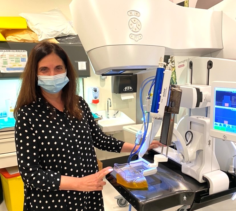
In a newly published study in the American Journal of Roentgenology, a team at Lawson Health Research Institute was the first in North America to find that a new breast cancer biopsy method may offer a more accurate and comfortable option for patients.
The method is a new form of mammography software that combines contrast enhanced mammography (CEM) with mammography guided biopsy technology at St. Joseph’s Health Care London’s Breast Care Program. These tools were combined in an effort to make the biopsy procedure more streamlined, accurate, and easier for patients and technicians.
CEM is a relatively new form of mammography that uses contrast iodine injected intravenously, which acts like a dye that allows radiologists to spot potential cancerous lesions more effectively. If potential lesions are found, a biopsy is often the next step.
Before this option was made available to patients through this research, suspicious lesion detection that was only seen on contrast enhanced mammography were biopsied under MRI. This meant longer procedures, and working with limited MRI availability.
“If a lesion is detected only by CEM we usually offer an MRI guided biopsy, but we first need to find the same lesion on an MRI,” says Dr. Anat Kornecki, Lawson Associate Scientist and Breast Radiologist Lead at St. Joseph’s Health Care London. “The problem is that it is sometimes hard to find the same lesion and the MRI itself can be uncomfortable for the patient. Also, some lesions that are close to implants or chest walls cannot be reached with MRI guided biopsy.”
Dr. Kornecki and her research team therefore decided to study this new method. They were the first in North America to trial CESM-guided biopsies by using new technology created by GE HeathCare. This software means that patients can have the biopsy done with the exact same modality, avoiding the need for an MRI.
The study included 50 patients through St. Joseph’s Breast Care Program. The research team found 51 potentially cancerous breast lesions. Biopsies were successfully performed for 46 of the lesions. The results showed that 11 were breast cancer, 10 were high-risk lesions, and the remaining were benign lesions.
“These are very similar results that were reported through MRI-guided biopsies, which means that this new method can replace the MRI,” explains Dr. Kornecki.
Patients also reported having a more comfortable experience with the CEM-guided biopsy method.
Researchers in London and at two other centres in Europe were the first to pilot this technique which has now been cleared by Health Canada and the FDA commercially. St. Joseph’s Breast Care Program was the first site in North America to offer this procedure as a clinical standard of care.
“It is a game changer with certainty,” adds Dr. Kornecki. “This is now a great added component for patients, which makes it a very good tool.”
Currently, CEM- guided biopsy can be offered to patients with lesions that were initially detected by MRI where a biopsy is not feasible due to the lesion location. While it is currently being used as a diagnostic tool only, Dr. Kornecki is hopeful that eventually CEM-guided biopsies will be approved as an initial breast cancer screening tool as well.
About Lawson Health Research Institute
Lawson Health Research Institute is one of Canada’s top hospital-based research institutes, tackling the most pressing challenges in health care. As the research institute of London Health Sciences Centre and St. Joseph’s Health Care London, our innovation happens where care is delivered. Lawson research teams are at the leading-edge of science with the goal of improving health and the delivery of care for patients. Working in partnership with Western University, our researchers are encouraged to pursue their curiosity, collaborate often and share their discoveries widely. Research conducted through Lawson makes a difference in the lives of patients, families and communities around the world. To learn more, visit www.lawsonresearch.ca.
Media Contacts
Celine Zadorsky
Senior Media Relations Consultant
Communications & Public Engagement
T: 519-685-8500 ext. 73502
Celine.zadorsky@lhsc.on.ca
Lawson study validates new biopsy method for breast cancer patients
In a newly published study in the American Journal of Roentgenology, a team at Lawson Health Research Institute was the first in North America to find that a new breast cancer biopsy method may offer a more accurate and comfortable option for patients.
The method is a new form of mammography software that combines contrast enhanced mammography (CEM) with mammography guided biopsy technology at St. Joseph’s Health Care London’s Breast Care Program. These tools were combined in an effort to make the biopsy procedure more streamlined, accurate, and easier for patients and technicians.
CEM is a relatively new form of mammography that uses contrast iodine injected intravenously, which acts like a dye that allows radiologists to spot potential cancerous lesions more effectively. If potential lesions are found, a biopsy is often the next step.
Before this option was made available to patients through this research, suspicious lesion detection that was only seen on contrast enhanced mammography were biopsied under MRI. This meant longer procedures, and working with limited MRI availability.
“If a lesion is detected only by CEM we usually offer an MRI guided biopsy, but we first need to find the same lesion on an MRI,” says Dr. Anat Kornecki, Lawson Associate Scientist and Breast Radiologist Lead at St. Joseph’s Health Care London. “The problem is that it is sometimes hard to find the same lesion and the MRI itself can be uncomfortable for the patient. Also, some lesions that are close to implants or chest walls cannot be reached with MRI guided biopsy.”
Dr. Kornecki and her research team therefore decided to study this new method. They were the first in North America to trial CESM-guided biopsies by using new technology created by GE HeathCare.This software means that patients can have the biopsy done with the exact same modality, avoiding the need for an MRI.
The study included 50 patients through St. Joseph’s Breast Care Program. The research team found 51 potentially cancerous breast lesions. Biopsies were successfully performed for 46 of the lesions. The results showed that 11 were breast cancer, 10 were high-risk lesions, and the remaining were benign lesions.
“These are very similar results that were reported through MRI-guided biopsies, which means that this new method can replace the MRI,” explains Dr. Kornecki.
Patients also reported having a more comfortable experience with the CEM-guided biopsy method.
Researchers in London and at two other centres in Europe were the first to pilot this technique which has now been cleared by Health Canada and the FDA commercially. St. Joseph’s Breast Care Program was the first site in North America to offer this procedure as a clinical standard of care.
“It is a game changer with certainty,” adds Dr. Kornecki. “This is now a great added component for patients, which makes it a very good tool.”
Currently, CEM- guided biopsy can be offered to patients with lesions that were initially detected by MRI where a biopsy is not feasible due to the lesion location. While it is currently being used as a diagnostic tool only, Dr. Kornecki is hopeful that eventually CEM-guided biopsies will be approved as an initial breast cancer screening tool as well.
Lawson's Top 12 Research Stories from 2020
Image

Image
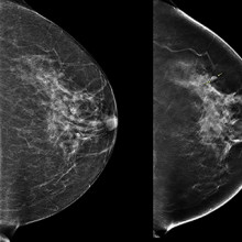
3D imaging technology could improve outcomes for patients with breast cancer
During a conventional digital 2D mammogram, two x-ray images are taken of the breast, one from top-to-bottom and another from side-to-side at an angle. This technology is limited by the overlapping breast tissue that occurs from the required compression of the breast, and breast abnormalities may be hidden. A study at Lawson is looking to determine if digital breast tomosynthesis, a type of 3D imaging, is better at detecting breast tissue abnormalities than the 2D mammography regularly used today. Read more.
Image
Image
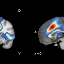
Antioxidants in the brain linked to improved treatment results in patients with psychosis
Once patients with psychosis start treatment, some get better in weeks while it can take months for others. A research team from Lawson and Western University studied antioxidant levels in the brain, and found that these chemicals, which rid the body of normal metabolic biproducts called free radicals, may improve outcomes of early intervention in psychosis. Read more.
Image
Image
Researchers awarded $4.8 million to validate locally developed test, EpiSign, for first-line diagnostic testing of rare hereditary disorders
A clinical trial named “EpiSign-CAN,” led by researchers at Lawson was awarded $4.8 million to measure the clinical impact of a new molecular genomics test for diagnosing genetic neurodevelopmental conditions. The diagnostic test, called EpiSign, uses machine learning to analyze the EpiSign Knowledge Database. This database compiles information on rare genetic diseases using laboratory analyses of the entire genome, referred to as the epigenome, from patients with suspected genetic abnormalities. Read more.
Image
Image

Perceptions of confidentiality for Canadian Veterans discussing moral injuries
Lawson researchers are exploring Canadian Veterans’ beliefs about confidentiality in mental health care and whether those beliefs act as a barrier to seeking treatment for a type of trauma known as moral injury. Moral injury describes psychological distress following events where a person performs, witnesses or fails to prevent acts that conflict with deeply held moral standards. Evidence suggests that moral injuries are on the rise among deployed members of the Canadian Armed Forces, and that those exposed to such events are at a higher risk of developing post-traumatic stress disorder and depression. Read more.
Image
Image
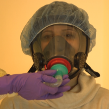
New device could reduce COVID-19 infection risk and demand for invasive ventilators
Researchers designed a non-invasive ventilation mask that could significantly reduce aerosolization – the production of airborne respiratory droplets that may contain viruses or bacteria – when treating patients with COVID-19. The new device aims to reduce infection risks associated with non-invasive ventilation and lessen the demand for invasive ventilators. It is currently being tested through a clinical trial with patients at London Health Sciences Centre (LHSC). Read more.
Image
Image
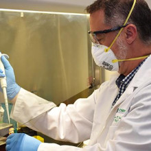
Researchers first in world to profile the body’s immune response to COVID-19
By studying blood samples from critically ill patients at LHSC, researchers identified a unique pattern of six molecules that could be used as therapeutic targets to treat COVID-19. Studies show that part of what makes the virus so deadly is that the body mounts an overreactive immune response as the virus grows and replicates. This response releases inflammatory molecules in order to fight the virus, but also destroys healthy cells and organs in the process. Read more.
Image
Image
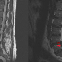
Study suggests that surgery may be superior treatment for chronic sciatica
In a randomized controlled trial, surgery was found to be superior to non-operative therapy in the treatment of chronic sciatica. Chronic sciatica can be caused by a disc herniation which compresses a nerve in the lumbar spine causing pain from the lower back to the leg. The primary treatment options for sciatica are surgery or non-operative care. Researchers conducted this study to test if a surgical treatment called microdiscectomy results in better patient outcomes for those with chronic sciatica compared to non-operative care. Read more.
Image
Image
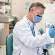
Researchers unravel two mysteries of COVID-19
A team from Lawson and Western University made significant steps forward in understanding COVID-19 through two back-to-back studies. In one study, the team identified six molecules that can be used as biomarkers to predict how severely ill a patient will become. In the other study, they were the first to reveal a mechanism causing blood clots in COVID-19 patients and potential ways to treat them. The studies were conducted by analyzing blood samples from critically ill patients at LHSC. Read more.
Image
Image
Gut microbiome may influence how cancer patients respond to oral therapies, study suggests
A study from Lawson and Western illustrated how the gut microbiome interacts with an oral medication in prostate cancer patients, suggesting bacteria in the gut play a role in treatment outcomes. The findings highlight how the drug abiraterone acetate is metabolized by bacteria in the gut to reduce harmful organisms while promoting those that fight cancer. The research team suspects this is one of many examples of how the microbiome influences our response to medications. Read more.
Image
Image
First Contrast Enhanced Spectral Mammography guided biopsy in North America
Researchers at Lawson performed the first breast biopsy guided by Contrast Enhanced Spectral Mammography (CESM) in North America on June 12, 2020. CESM is a novel diagnostic imaging tool that is able to detect cancerous lesions at a greater rate than standard mammography, and at close rate to MRI. The procedure is faster and more accurate, comfortable and cost effective than an MRI biopsy. Read more.
Image
Image

Assessing the pandemic's impact on Canadian Veterans and their spouses
A project from Lawson and the Centre of Excellence on Post-Traumatic Stress Disorder (PTSD) hopes to discover the impact the COVID-19 pandemic is having on the mental health of Canadian Veterans and their spouses. They are partnering with up to 1,000 Canadian Veterans and 250 spouses of Canadian Veterans. Through online surveys, the project will hear directly from Veterans and their spouses to assess the pandemic’s effects on their well-being over time. Read more.
Image
Image
Fecal transplants show promise as treatment for non-alcoholic fatty liver disease
A randomized controlled trial found that fecal transplants in patients with non-alcoholic fatty liver disease (NAFLD) results in a reduction in how easily pathogens and other unwanted molecules pass through the human gut and into circulation, known as intestinal permeability. The results could have implications for the treatment of numerous conditions including metabolic syndrome and autoimmune diseases. Read more.
… link to best in breast care conference 2018 flyer …
… Managing Breast Pain …
… 2 adult females diagnosed with paranoid schizophrenia and breast 5 Book of Abstracts RMHC 9th Annual Research Half-Day … 2 adult females diagnosed with paranoid schizophrenia and breast 5 Book of Abstracts RMHC 9th Annual Research Half-Day …
Regional Mental Health Care London and St. Thomas 9th Annual Research Half Day, May 14, 2008 BOOK OF ABSTRACTS & RESEARCH REPORT 2007 by RMHC RESEARCH COMMITTEE 1 Book of Abstracts RMHC 9th Annual Research Half-Day May 14, 2008 and Research Report 2007 RESEARCH INSIGHTS of the Regional Mental Health...
… Residency Program Director. James Calvin Narinder Paul Body Breast Cardiothoracic Musculoskeletal Neuroradiology … Residency Program Director. James Calvin Narinder Paul Body Breast Cardiothoracic Musculoskeletal Neuroradiology …
GUIDELINES FOR PHYSICIAN PERFORMANCE MANAGEMENT - Listing of Physician Leaders in the Department/Organization DEPARTMENT DEPARTMENT CHIEFS SITE CHIEFS DIVISION DIVISION CHIEFS/SECTION HEADS/ DIVISION CHAIRS/MEDICAL DIRECTORS OTHER LEADERSHIP ROLE RESIDENCY PROGRAM DIRECTORS COMMENTS Ashraf Fayad (LH...
… including: Body Breast Cardiothoracic MSK Neurological … including: Body Breast Cardiothoracic MSK Neurological …
ApprovedbyDepartmentChief,CWCCandMACJanuary2019 Page1 Delineation of Procedural Privileges Department of Medical Imaging Division of Diagnostic Radiology All members of the Division of Diagnostic Radiology must have their Fellowship in Diagnostic Radiology and accreditation in their appropriate su...
… specialized in Diagnostic Radiology, including: Body Breast Cardiothoracic MSK Neurological Pediatrics … specialized in Diagnostic Radiology, including: Body Breast Cardiothoracic MSK Neurological Pediatrics …
Approved by Department Chief, CWCC and MAC January 2019 Page 1 Delineation of Procedural Privileges Department of Medical Imaging Division of Diagnostic Radiology All members of the Division of Diagnostic Radiology must have their Fellowship in Diagnostic Radiology and accreditation in their appro...
m ST::JOSEPH'S A publication of St. Josephs Health Care London WINTER 2020 | ISSUE 01 BORN TO MOVE St. Josephs leads the way for those facing mobility challenges with the creation of The Gray Centre for Mobility and Activity. 1 201 ISSUE www.sjhc.london.on.ca ~ ~ OSEPH'S 11 C ~~ S!JOSEPHs HEALTH CAR...
… condition are more common than heart attack, stroke and breast cancer combined. The fallout can be debilitating, … heart who suffer a hip fracture will die attack, stroke and breast within the following year. cancer combined. 1 IN 3 … of a tiny dot with big implications for women with dense breasts. It was startlingly clear – a tiny bright dot stood …
Our entrances have m S1: A publication of St. Joseph s Health Care London WINTER 2021 | ISSUE 03 Our entrances have KEEPING IT SAFE become a gateway of safety to protect our patients and sta . It's a responsibility more than 160 screeners take very seriously. These "perimeter protectors" are vital ...
… YOur guide to Breast assessment Breast Care Program St. Joseph’s Hospital 268 Grosvenor St. London, ON N6A 4V2 THE BEST IN BREAST CARE The Breast Care Program at St. Joseph’s Health … YOur guide to Breast assessment Breast Care Program St. Joseph’s Hospital 268 Grosvenor St. …
YOur guide to Breast assessment Breast Care Program St. Josephs Hospital 268 Grosvenor St. London, ON N6A 4V2 THE BEST IN BREAST CARE The Breast Care Program at St. Josephs Health Care London provides a confidential, coordinated, specialized breast assessment service. The goal of the program is to m...
… have any of the following? Colon Year: Lung Year: Breast Year: Cervical Year: Prostate Year: Connective … have any of the following? Colon Year: Lung Year: Breast Year: Cervical Year: Prostate Year: Connective …
Date: Why are you seeing the Doctor today? What is your marital status (circle)? Single Married Common-law Widowed Divorced Separated What is your age? ____________ How many children do you have? What is your Occupation? Are you on Disability? YES NO What is your Drug Plan: Private Insurance Over...
Over $2 million in federal funding to advance discoveries in health research
Last week, the Honourable Kirsty Duncan, Minister of Science and Sport, announced an unprecedented investment of more than $588 million through the Natural Sciences and Engineering Research Council of Canada’s (NSERC) Discovery Grants program.
The successful applications in London include 12 projects funded for Lawson Health Research Institute scientists, through Western University. In total, they will receive $2.3 million in funding over five years.
“The funding demonstrates our strong and enduring commitment to science and researchers. Since taking office, our government has worked hard to bring science and research back to their rightful place and this historic investment in the discoveries of tomorrow is just one example of how we are achieving this goal,” says The Honourable Kirsty Duncan, Minister of Science and Sport.
Across Canada, this funding will go to more than 4,850 researchers and students as they pursue their world-leading discovery work. It also includes support for nearly 500 early-career researchers who will bring a diversity of new voices and new insights to their fields.
Local research highlights
Dr. Jeffrey Carson is exploring the role of photoacoustic imaging as a method for detecting breast cancer. Currently, during the procedure the breast must be submerged in a tank of water to enhance the transmission of photoacoustic waves from the breast to the sensors.
“The water tank is cumbersome and impractical,” notes Dr. Carson. “Our goal is to eliminate the need for the water tank by detecting the photoacoustic waves through the air without making contact with the breast.” Their hope is that the project leads to the development of a non-contact photoacoustic scanner offering women the opportunity to sit or stand comfortably during breast imaging. “This simple improvement could greatly accelerate the adoption of photoacoustic imaging for breast cancer screening.”
Dr. Carson adds that funding like this provides graduate student trainees opportunities to develop knowledge and skills in engineering, mathematics, and biophysics. “They bring innovative new ideas to the Canadian medical device industry through employment and entrepreneurship.”
Image

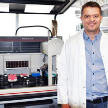
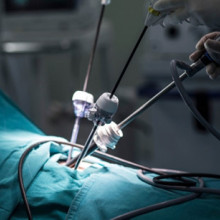
Dr. Rajnikant Patel is developing advanced robotic and intelligent systems for the next generation of systems and devices for minimally invasive surgery and therapy. These reduce trauma and costs while enhancing efficiency and reliability.
“For us, this funding opportunity is unique because it supports a program of research rather than a project,” says Dr. Patel. “We can explore new areas and directions that will lead to research projects and medical applications. A program that investigates novel robotic and AI technologies fits well with NSERC’s mandate.”
Image
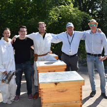
Dr. Gregor Reid is leading a revolutionary project that could save the world’s honeys bees, insects that are vital to human survival.
The intent is to develop an understanding of how lactobacilli strains can counter the most widely used pesticides that are wiping out nature's critically important pollinators. The lactobacilli appear to potentially degrade some of these toxic chemicals and improve the ability of honey bees to fight off early death.
“NSERC funding gives ideas like this a chance and even though the funding amount is relatively small, it allows graduate students to apply for their own awards and work on the project,” explains Dr. Reid. Students Brendan Daisley and Johnny Chmiel have been awarded NSERC scholarships to work on this important research.
Image
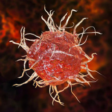
Dr. Xiugen Zheng’s project is investigating the role of circular RNA AEBP in the development and function of dendritic cells which are very important immune cells in the immune system. This will provide insights into new molecular and gene regulators, and their impact on the immune system and overall health.
“NSERC research funding greatly supports us to study the basic scientific questions that are critical for better understanding health problems and developing treatment for diseases,” says Dr. Zheng.
Image
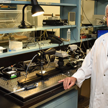
Dr. Rudolf Veldhuizen’s laboratory aims to understand how surfactant performs its function at a molecular and biophysical level. Pulmonary surfactant is a material in the lung that allows people and other mammals to breath with minimal effort. You can see this in babies who are born prematurely and have trouble breathing due to the lack of surfactant.
“Previous work has established a generalized model of how surfactant improves lung function under standardized conditions. This, however, does not explain how surfactant functions in extreme conditions,” says Dr. Veldhuizen. “By exploring conditions in comparative, mechanistic studies we will be able to establish a more universal understanding of surfactant function.”
This funded work provides a foundation for clinically relevant studies to further explore the role of alterations to surfactant in lung injuries and the opportunities for therapeutic interventions.
Image
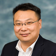
Dr. Shou Li is developing state-of-the-art machine learning system able to analyze huge amounts of clinical data and provide human level intelligent analysis. This work will enable the prediction of disease onset, progression and prognosis. “It is added value that will lead to more effective and efficient health care,” explains Dr. Li.
He adds that this funding is supporting a multi-disciplinary research program that combines the strength of multiple teams. “We will look at both the fundamental side of machine learning systems and clinical applications. In this way, we connect basic science with clinical science.”
Image
Congratulations to all the Lawson scientists who received funding:
- Dr. Dean Betts for Metabolic reprogramming to enhance the generation of canine induced pluripotent stem cells (Physiology and Pharmacology)
- Dr. Jeffrey Carson for Development of non-contact photoacoustic tomography (Medical Biophysics)
- Dr. Louis Ferreira for Multi-Directional Mechanical Testing of Bone using CTCompatible Loading Mechanisms (Mechanical and Materials Engineering)
- Dr. Shuo Li for Innovative Machine Learning for Medical Data Analytics (Medical Imaging)
- Dr. Penny MacDonald for Investigating cognitive functions mediated by ventral and dorsal striatum (Clinical Neurological Sciences)
- Dr. Charles McKenzie for Fetoplacental Molecular and Metabolic Magnetic Resonance Imaging; instalment (Medical Biophysics)
- Dr. Rajnikant Patel for Design and Control of Robotic Systems and Devices for Medical Applications (Electrical and Computer Engineering)
- Dr. Gregor Reid for Detoxification functionality of lactic acid bacteria (Microbiology and Immunology)
- Dr. Rudolf Veldhuizen for Mechanisms of surface tension reduction by pulmonary surfactant (Physiology and Pharmacology)
- Dr. Aaron Ward for Machine learning-based quantitative image, tissue, and clinical data analysis for lesion detection and characterization on prostate cancer imaging (Medical Biophysics)
- Dr. Eugene Wong for Optimization of spatiotemporal-modulated electric fields and fabrication of organs-on-chips for applications in Medical Physics (Physics and Astronomy)
- Dr. Xiufen Zheng for The role of circular RNA AEBP2 in dendritic cells (Pathology)
Scientist
… Prostheses and Supports • external prostheses. Charges for breast prostheses are subject to a maximum of 6 per Benefit … Prostheses and Supports • external prostheses. Charges for breast prostheses are subject to a maximum of 6 per Benefit …
Council of Academic Hospitals of Ontario (CAHO) Plan Document Number: G0086936 Plan F: London Health Science Centre - Plan A Employee Name: Certificate Number: Welcome to Your Group Benefit Program Plan Document Effective Date: January 1, 2012 This Benefit Booklet has been specifically designed with...
… Prostheses and Supports external prostheses. Charges for breast prostheses are subject to a maximum of 6 per Benefit … Prostheses and Supports external prostheses. Charges for breast prostheses are subject to a maximum of 6 per Benefit …
Council of Academic Hospitals of Ontario (CAHO) 1 Council of Academic Hospitals of Ontario (CAHO) Plan Document Number: G0086936 Plan F: London Health Science Centre - Plan A Employee Name: Certificate Number: Welcome to Your Group Benefit Program Plan Document Effective Date: January 1, 2012 This B...
… Prostheses and Supports • external prostheses. Charges for breast prostheses are subject to a maximum of 6 per Benefit … Prostheses and Supports • external prostheses. Charges for breast prostheses are subject to a maximum of 6 per Benefit …
Council of Academic Hospitals of Ontario (CAHO) 1 Council of Academic Hospitals of Ontario (CAHO) Plan Document Number: G0086936 Plan F: London Health Science Centre - Plan A Employee Name: Certificate Number: Welcome to Your Group Benefit Program Plan Document Effective Date: January 1, 2012 This B...
… Prostheses and Supports external prostheses. Charges for breast prostheses are subject to a maximum of 6 per Benefit … Prostheses and Supports external prostheses. Charges for breast prostheses are subject to a maximum of 6 per Benefit …
Council of Academic Hospitals of Ontario (CAHO) Plan Document Number: G0086936 Plan F: London Health Science Centre - Plan A Employee Name: Certificate Number: Welcome to Your Group Benefit Program Plan Document Effective Date: January 1, 2012 This Benefit Booklet has been specifically designed with...
… St. Joseph’s Hospital Breast Care Program, Room D1-112 268 Grosvenor St., P.O. Box … you provide on the Patient History form will be used by the Breast Care Program surgeon and clinic staff to plan and … at the bottom of page 2 to submit the form to St. Joseph’s Breast Care Program by e-mail. If the button does not link …
St. Josephs Hospital Breast Care Program, Room D1-112 268 Grosvenor St., P.O. Box 5777, Stn. B London, ON N6A 4V2 Tel. 519-646-6000 ext. 65020 Patient History Form Instructions The information you provide on the Patient History form will be used by the Breast Care Program surgeon and clinic staff to...