Search
Search
251 Search Results:
… Patients Guide to Breast Lumpectomy …
… Director. James Calvin Stephen Wetmore Narinder Paul Body Breast Cardiothoracic Musculoskeletal Neuroradiology … Director. James Calvin Stephen Wetmore Narinder Paul Body Breast Cardiothoracic Musculoskeletal Neuroradiology …
GUIDELINES FOR PHYSICIAN PERFORMANCE MANAGEMENT - Listing of Physician Leaders in the Department/Organization DEPARTMENT DEPARTMENT CHIEFS SITE CHIEFS DIVISION DIVISION CHIEFS/SECTION HEADS/ DIVISION CHAIRS/MEDICAL DIRECTORS OTHER LEADERSHIP ROLE RESIDENCY PROGRAM DIRECTORS COMMENTS Ashraf Fayad (LH...
… - UH) Ian Ross (LHSC - VH) Justin Amann (St. Joseph's) Body Breast Cardiothoracic Musculoskeletal Neuroradiology … - UH) Ian Ross (LHSC - VH) Justin Amann (St. Joseph's) Body Breast Cardiothoracic Musculoskeletal Neuroradiology …
GUIDELINES FOR PHYSICIAN PERFORMANCE MANAGEMENT - Listing of Physician Leaders in the Department/Organization DEPARTMENT DEPARTMENT CHIEFS SITE CHIEFS DIVISION DIVISION CHIEFS/SECTION HEADS/ DIVISION CHAIRS/MEDICAL DIRECTORS OTHER LEADERSHIP ROLE RESIDENCY PROGRAM DIRECTORS COMMENTS ANESTHESIA & PER...
… Director. James Calvin Stephen Wetmore Narinder Paul Body Breast Cardiothoracic Musculoskeletal Neuroradiology … Director. James Calvin Stephen Wetmore Narinder Paul Body Breast Cardiothoracic Musculoskeletal Neuroradiology …
GUIDELINES FOR PHYSICIAN PERFORMANCE MANAGEMENT - Listing of Physician Leaders in the Department/Organization DEPARTMENT DEPARTMENT CHIEFS SITE CHIEFS DIVISION DIVISION CHIEFS/SECTION HEADS/ DIVISION CHAIRS/MEDICAL DIRECTORS OTHER LEADERSHIP ROLE RESIDENCY PROGRAM DIRECTORS COMMENTS Ashraf Fayad (LH...
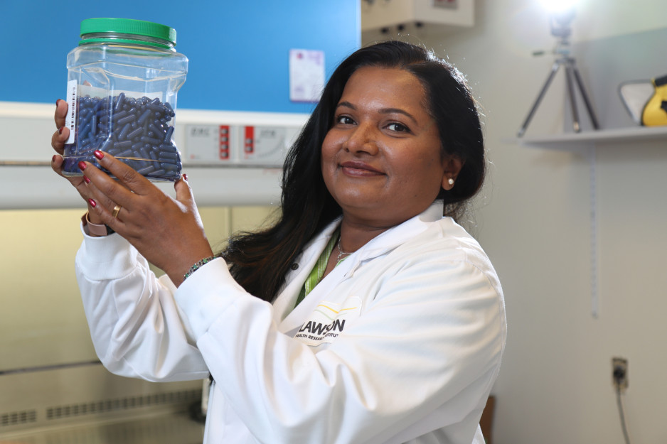
Poop in a pill helping advance cancer care
Lawson Research Institute scientists have perfected the delivery of fecal transplants via patient-friendly capsules now central in ground-breaking cancer treatment studies.
It’s one of the most exciting areas of research in cancer care.
Description
Making waves in scientific and health care circles worldwide, it holds the potent potential to “jazz up” cells that attack cancer and boost the body’s response to treatment.
So what is this powerful ally? It’s poop in a pill – home-grown right here at St. Joseph’s Health Care London and Lawson Research Institute.
In fact, Lawson scientists Dr. Michael Silverman, Seema Nair Parvathy, PhD and their team are considered poop pill pioneers, having perfected the delivery of fecal transplantation by way of patient-friendly capsules that can be easily swallowed. These capsules contain healthy gut microbes that have become pivotal in many landmark cancer treatment studies.
Triggering an immune response
Understanding the role of poop in cancer treatment requires grasping the wonders of the human microbiome and its key role in influencing health and well-being.
The human microbiome consists of trillions of microorganisms that live inside and outside of the body, including bacteria, viruses and yeasts. While some bacteria are associated with disease, others are vital to the human immune system – the body’s main protective and disease-fighting tool – and many other aspects of health. Over the past decade, microbiome research has led to a revolution in medicine as scientists unravel just how an imbalance of these microorganisms interferes with many aspects of good health.
"(Fecal microbial transplants) allows us to harness the immune system to mount a stronger defence." Dr. Michael Silverman
The goal of fecal microbiota transplants (FMT) is to transfer healthy gut microbes from donors into patients with cancer and other diseases so that healthy bacteria will colonize in the patient’s gut and improve the microbiome, explains Silverman, Medical Director of St. Joseph’s Infectious Diseases Care Program and citywide Chief of Infectious Diseases for London’s hospitals.

To do so, stools are collected from carefully screened healthy donors, prepared in a lab into capsule format, and introduced into a patient’s gastrointestinal tract.
“What is so exciting when it comes to cancer treatment is the evidence we now have showing how a healthy microbiome activates the immune response to tumours to make the treatment more effective,” Silverman adds. “It allows us to harness the immune system to mount a stronger defense.”
St. Joseph’s capsules are central to several significant studies currently underway aimed at improving treatment for lung, kidney, breast, renal, pancreatic and other cancers.
Among the most notable is the London team’s lead role in a ground-breaking national study – one of the world’s largest randomized controlled clinical trials using FMT to improve the effectiveness of the standard of care for advanced melanoma, a type of skin cancer.
Improving melanoma survival rates
About 11,300 Canadians will be diagnosed with melanoma in 2024 and, even with standard treatment, about half that number will experience disease progression and die.
The 16-site Canadian trial builds off the work of Silverman, Parvathy and their team, in partnership with Saman Maleki, PhD, and Dr. John Lenehan at London Health Sciences Centre. Together, they were the first to demonstrate the safety and therapeutic potential of using the capsules produced at St Joseph’s to influence a patient’s gut microbiota to enhance immunotherapy and increase the odds of surviving advanced melanoma.
“London is seen as having the most expertise in use of FMT in cancer care in the world and is a driving force in moving this forward,” says Silverman. “Immunotherapy is rapidly expanding the number of treatable cancers and our FMT therapy is helping to accelerate this progress.”
… Clients making a difference 16 Women find support through breast assessment program 17 Partnerships provide support to … Centre Acute/Ambulatory Care • 3M Osteoporosis Clinic • Breast Centre • Centre for Lung Health and Home Oxygen … estimated that 21,600 Canadian women will be diagnosed with breast cancer and 5,300 will die of it. Every week across …
S T. J O S E P H S H E A LT H C A R E , LO N D O N 2 0 0 4 / 2 0 0 5 care is at our core STAND TALL AND PROUD SINK YOUR ROOTS DEEPLY INTO THE EARTH REFLECT THE LIGHT OF A GREATER SOURCE THINK LONG TERM GO OUT ON A LIMB REMEMBER YOUR PLACE AMONG ALL LIVING BEINGS EMBRACE WITH JOY THE CHANGING SEASON...
… (please print): PHYSICIAN SIGNATURE: Phone Number: Fax: **BREAST ASSESSMENT FORM MUST BE FILLED OUT FOR ALL BREAST ULTRASOUNDS AND FAXED TO ST. JOSEPH’S ** August 17, … (please print): PHYSICIAN SIGNATURE: Phone Number: Fax: **BREAST ASSESSMENT FORM MUST BE FILLED OUT FOR ALL BREAST …
ULTRASOUND REQUISITION Site: London Health Sciences Centre Vic/Childrens F: 519-667-6826 St. Josephs Health Care London F: 519-646-6204 London Health Sciences Centre UH F: 519-633-3034 PATIENT INFORMATION: Surname: First Name: Middle Initial: Gender: Date of Birth (YYYY-MM-DD): Street Address: Apa...
… • St. Joseph’s Hospital is designated as an official breast assessment centre affiliated with the Ontario 27 Breast Screening Program • Armatec Survivability Corporation … • St. Joseph’s Hospital is designated as an official breast assessment centre affiliated with the Ontario 27 …
ST. JOSEPHS HEALTH CARE LONDON | ANNUAL REPORT TO THE COMMUNITY | 2002/2003 2 Dedication This years Annual Report to the Community is dedicated to the St. Josephs Health Care, London staff members and volunteers who continue to give so much of themselves to help manage SARS restrictions. Thank you t...
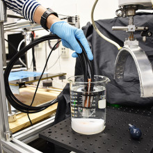
Researchers developing photoacoustic hand-held probe for tumour detection during breast conserving surgery
Researchers at Lawson Health Research Institute (Lawson) are developing a hand-held photoacoustic imaging probe to be used during breast conserving surgery to quickly and accurately verify if all cancerous tissue has been removed.
Surgeons currently do not have real-time technology to guide tumour removal during surgery.
Using current tools, there is a 20 per cent chance that cancerous cells will be left behind, risking recurrence and repeat surgery.
Breast cancer represents 25 per cent of all new cancer diagnoses in women and 13 per cent of all cancer related deaths in women. Treatment for breast cancer often requires either complete breast removal in severe cases, or surgical removal of the cancerous tumour in combination with other therapies. Removing only the tumour is called breast conserving surgery.
Image

Image
Photoacoustic Screening
The new device is an extension of the photoacoustic screening (iPAS) technology developed in the laboratory of Dr. Jeffrey Carson, Principal Investigator and Lawson Scientist. The technique uses light and sound to capture 3D images of surgically removed breast tissue. Their studies show that iPAS can catch up to 75 per cent of missed tumour cells, decreasing the odds of failed surgery to five per cent.
Dr. Muriel Brackstone, Associate Scientist at Lawson, Head of the Breast Care Clinic at St. Joseph’s Hospital London, and Surgical Oncologist at London Health Sciences Centre, brings her clinical expertise to the project.
“With the first generation iPAS technology, we would remove the tumour, take it to the lab for imaging and wait to see if there was a rim of normal tissue around the removed tumour so we knew it was removed completely. The wait was anywhere from 20 minutes to an hour. During that time, the patient is under anesthesia, the surgical team is idle and precious OR time is being used,” explains Dr. Brackstone.
A hand-held tool that surgeons can use
Creation of a hand-held probe to be used in the operating room is the next step in the advancement of this new technology. Elina Rascevska, biomedical engineering student at Western University, recently joined the Lawson team to convert lab-based iPAS technology into a hand-held device.
“We have developed a prototype of the iPAS probe, and once we can verify the quality of the images it produces, we will give it to Dr. Brackstone to test in the OR,” says Rascevska.
The iPAS probe does not need a trained operator and would be used by the surgical team. Instead of imaging the removed tissue, it scans the surgical cavity in real time to give the team a faster and more accurate indication as to whether the cancerous tissue has been removed.
“If we can progress this technology to a point where physicians can use it as part of standard protocols, we will have reduced the amount of time each patient needs to spend in the OR, the amount of call-backs and repeat surgeries, and ultimately improve quality of life for patients with breast cancer,” adds Dr. Carson.
Image
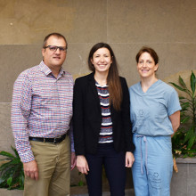
(From left): Dr. Jeffrey Carson, Elina Rascevska, Dr. Muriel Brackstone
Scientist
… months Central Nervous System CT Scan Head 2 mSv 8 months Breast Mammography 0.7 mSv 3 months Pelvis (Hip) Bone … months Central Nervous System CT Scan Head 2 mSv 8 months Breast Mammography 0.7 mSv 3 months Pelvis (Hip) Bone …
CARING FOR THE BODY, MIND & SPIRIT SINCE 1869 Renowned for compassionate care, St. Josephs is one of the best academic health care organizations in Canada dedicated to helping people live to their fullest by minimizing the effects of injury, disease and disability through excellence in care, teachin...
… (low in Salicylates) Ingredients • 3 cups cooked chicken breast, cubed • 2 cups golden delicious apple, peeled and … (low in Salicylates) Ingredients • 2 lbs boneless chicken breast • 2 TBL lime juice • 1 onion, chopped (optional) • 1 … Ingredients • 5 potatoes, sliced and peeled • 4 chicken breasts • parsley pesto (see recipe) Directions Cover …
The Salicylate Sensitivity Cookbook The Salicylate Sensitivity Cookbook The Salicylate Sensitivity Cookbook Starting a new restricted diet can be daunting. It is easy to get into a rut and make the same safe meals over and over again. The aim of this cookbook is provide inspiration and ideas ...
… and teaching around the world; the Norton and Lucille Wolf Breast Care Centre is a unique ambulatory clinic model where … assessment, diagnostics and surgical care and advances breast care teaching and research; The Ivey Eye Institute is … and teaching around the world; the Norton and Lucille Wolf Breast Care Centre is a unique ambulatory clinic model where …
STRATEGIC PLAN 2015 | 2018ST. JOSEPHS HEALTH CARE LONDON Realizing our Vision St. Josephs Vision, Mission and Values OUR VISION OUR MISSION OUR VALUES RESPECT, EXCELLENCE, COMPASSION STRATEGIC PLAN 2015 TO 2018 Our Patients, Residents and Families GOAL GUIDED BY THEIR VOICES; EXCELLENCE ALWAYS STRA...
… 2018-2019 and will focus on patients served by St. Joseph’s Breast Care Program. Engagement of Clinicians, Leadership & … 2018-2019 and will focus on patients served by St. Joseph’s Breast Care Program. Engagement of Clinicians, Leadership & …
St. Josephs Health Care London 1 PO Box 5777, STN B London, ON N6A 4V2 Quality Improvement Plan (QIP) Narrative for Health Care Organizations in Ontario 3/29/2018 This document is intended to provide health care organizations in Ontario with guidance as to how they can develop a Quality Improvement ...
… and teaching around the world; the Norton and Lucille Wolf Breast Care Centre is a unique ambulatory clinic model where … assessment, diagnostics and surgical care and advances breast care teaching and research; The Ivey Eye Institute is … and teaching around the world; the Norton and Lucille Wolf Breast Care Centre is a unique ambulatory clinic model where …
STRATEGIC PLAN 2015 | 2018ST. JOSEPHS HEALTH CARE LONDON Realizing our Vision St. Josephs Vision, Mission and Values OUR VISION OUR MISSION OUR VALUES RESPECT, EXCELLENCE, COMPASSION STRATEGIC PLAN 2015 TO 2018 Our Patients, Residents and Families GOAL GUIDED BY THEIR VOICES; EXCELLENCE ALWAYS STRA...
… • We will have implemented a patient portal pilot in the Breast Care Centre and developed a plan for further patient … • We will have implemented a patient portal pilot in the Breast Care Centre and developed a plan for further patient …
sjhc.london.on.ca 2018-2021 Strategic Plan Reaching Out Connecting Care Innovating Together Contents Board Chair and CEO Message Background Mission, Vision, Values Strategic Priorities Reaching Out Connecting Care Innovating Together Leveraging Technology Empowering People Key Principles Uncom...