Search
Search
2802 Search Results:
… Department of Medical Affairs Standard Operating Procedure HR Planning and Credentialing … No: 001 Issued By: HR Planning and Credentialing Specialist Supersedes: Approved By: Integrated Director, Medical … 12, 2022 1. PURPOSE The attached document appended to this Standard Operating Procedure (SOP) titled “HR Planning and …
Department of Medical Affairs Standard Operating Procedure HR Planning and Credentialing Guide for Clinical Departments Section: [Credentialing] SOP No: [A-1-001] Section: CREDPS SOP No: 001 Issued By: HR Planning and Credentialing Specialist Supersedes: Approved By: Integrated Director, Medical Aff...
… June 2012 Ongoing Engagement As part of St. Joseph’s commitment to supporting our patients, staff and visitors, Southwest Centre is a designated smoke-free facility, which … June 2012 Ongoing Engagement As part of St. Joseph’s commitment to supporting our patients, staff …
June 2012 Ongoing Engagement As part of St. Josephs commitment to supporting our patients, staff and visitors, Southwest Centre is a designated smoke-free facility, which includes the building and the grounds. Below are some common questions and answers. Q - Why is the Southwest Centre for Forensic ...
… kidney function (eGFR value of less than 30). Preparation Instructions Please follow the instructions in this handout very carefully. The instructions have been tested to ensure they minimize … kidney function (eGFR value of less than 30). Preparation Instructions Please follow the instructions in this handout …
CT Colonography Alternate Bowel Prep This preparation is for patients with impaired kidney function (eGFR value of less than 30). Preparation Instructions Please follow the instructions in this handout very carefully. The instructions have been tested to ensure they minimize discomfort while produci...
… disease (eGFR value of less than 30), have your doctor call St. Joseph’s Medical Imaging department for alternate preparation instructions. Preparation Instructions Please follow the instructions in this handout … disease (eGFR value of less than 30), have your doctor call St. Joseph’s Medical Imaging department for alternate …
CT Colonography Bowel Prep This preparation is for patients with normal or mildly impaired kidney function (eGFR value of 30 or greater). If you have serious kidney disease (eGFR value of less than 30), have your doctor call St. Josephs Medical Imaging department for alternate preparation instructio...
… F: 519-524-8532 ☐ Middlesex Hospital Alliance - Strathroy F: 519-246-5930 ☐ Grey Bruce Health Services - … Health Centre -Walkerton F: 519-881-1388 ☐ Hanover and District Hospital F: 519-364-0062 ☐ St. Joseph's Health Care London F: 519-646-6204 ☐ Huron … F: 519-524-8532 ☐ Middlesex Hospital Alliance - Strathroy F: 519-246-5930 ☐ Grey Bruce Health Services - …
CT REQUISITION this form can be found on www.swpca Check one Site: Alexandra Marine and General Hospital-Goderich F: 519-524-8532 Middlesex Hospital Alliance - Strathroy F: 519-246-5930 Grey Bruce Health Services - Owen Sound F: 519-376-3952 South Bruce Grey Health Centre -Walkerton F: 519-881-...
… MIND & SPIRIT SINCE 1869 Renowned for compassionate care, St. Joseph’s is one of the best academic health care organizations in Canada dedicated to helping people live to their fullest by minimizing the effects of injury, disease and … MIND & SPIRIT SINCE 1869 Renowned for compassionate care, St. Joseph’s is one of the best academic health care …
CARING FOR THE BODY, MIND & SPIRIT SINCE 1869 Renowned for compassionate care, St. Josephs is one of the best academic health care organizations in Canada dedicated to helping people live to their fullest by minimizing the effects of injury, disease and disability through excellence in care, teachin...
Cultivating ‘eureka’ moments
Discovery should be ‘everyone, everywhere,’ says Lawson Research Institute Scientific Director Lisa Porter.
Lisa Porter believes excellence in health research is a continuum of inquiry, inspiration, innovation and improved patient care.
That’s why she is so energized by the promise and potential of Lawson Research Institute (Lawson) at St. Joseph’s Health Care London (St. Joseph’s), where she is Scientific Director and Vice President Research.
“Discovery comes from exploring great questions. You can’t have a ‘eureka’ moment without asking why things work, or don’t work – and that’s what we do so well here at St. Joseph’s,” says Porter.

A distinguished scientist herself with a passion for asking those probing questions, Porter leads strategic planning for research across the organization. Her vision includes growing the rich culture of research in several specialty pillars, while also reinforcing direct links between scientific inquiry and patient health.
“There’s data to show that patients who are treated in research-intensive hospitals live longer. That’s not just patients in clinical trials who benefit; that’s all patients who live longer,” she notes.
Other elements of her vision for Lawson include elevating data sharing and research support, expanding training opportunities for young researchers, growing grant support, strengthening collaboration and partnerships, and building relationships and reputation.
“I love that excellence is one of the values of St. Joseph’s. Excellence doesn’t mean we have all the answers. It means we’re continuously striving to be better. It means we’re asking questions that can drive better health care – not just for the patients we serve, but for national and global impact, too.”
Porter comes from a family of knowledge-seekers and problem-solvers. Her father repaired electronics and was an avid inventor. Her mother was a self-taught income –tax preparer with meticulous attention to detail. They ignited in her a curiosity that continued through her undergraduate studies in biology and pharmacology, her graduate and postdoctoral work, and her research as a cancer scientist at University of Windsor and founding director of its WE-SPARK Health Research Institute.
Now at Lawson, she wants to encourage, inspire and spotlight the innovative work of researchers, scientists, clinicians and students who are passionate about improving health.
“I want research to be everyone, everywhere,” she says. “We need hospitals, industry, people with lived experience, and policy makers coming into the fray. It can’t be just the researcher, the scientist. It’s about having champions embedded in all walks of life, from first line of care to people who can influence systemic change. It’s a messy piece, but it’s also how we fulfil this bigger mission to help everyone who comes to us for health care.”
Current Participants
Thank you for volunteering as a clinical research participant at HULC. You are helping to contribute to our growing knowledge and the advancement of clinical care. Please use these resources to assist in your role as a research participant.
Getting Here
The Roth | McFarlane Hand & Upper Limb Centre (HULC) is located at St. Joseph’s Hospital.
St. Joseph’s Hospital
Room D0-101
268 Grosvenor Street
London, Ontario N6A 4V2 519 646-6100 ext. 64640
Find turn-by-turn directions to HULC.
Contact Us
If you have any questions related to the research study you’re participating in, please contact the HULC clinical research team at 519-646-6100 ext. 64640.
Cutting Edge: Surgical advancement through research
Before the bright lights of the operating room are turned on and the surgeons and operating room staff are gowned and ready, research conducted at Lawson Health Research Institute has backed many of the surgical innovations and firsts performed at London Health Sciences Centre and St. Joseph’s Health Care London.
On October 5, Lawson hosted a Café Scientifique event where a panel of surgeons who are also Lawson scientists discussed their cutting-edge work. Guests had the opportunity to ask questions as part of an open-forum discussion to gain insights from the speakers, and from one another.
In celebration of Canada’s 150th anniversary as a nation, this event was the second of a two-part series focusing on the future vision for health care in Canada and the legacy that research at Lawson will leave. Research and knowledge-creation have been a hallmark of the various surgical areas at LHSC and St. Joseph’s since their inception, and the relationship between innovation and improving patient care has been an enduring trademark. Surgeon scientists have conducted and published research that has changed clinical practice worldwide.
Hand surgery: How small advances turn into complex surgical achievements
Dr. Bing Siang Gan, Lawson scientist, plastic surgeon, Hand and Upper Limb Centre, St. Joseph's
Image
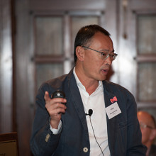
Dr. Gan has a particular research interest in the biology and treatment of Dupuytren's contracture and he uses conventional as well as minimal invasive procedures such as needle aponeurotomy and new collagenase enzyme injections to treat patients.
Dr. Gan explained how a better surgical understanding of Dupuytren's contracture combined with an understanding of the underlying gene factors, DNA, RNA, proteins, receptors, and collagen formation of the condition has led to pharmacological treatment options. The next step will be developing treatment options at every stage of Dupuytren's contracture to keep patients away from the operating room.
Transplant organ preservation: The best option may be “Stinky”
Dr. Alp Sener, Lawson scientist, transplant surgeon, Multi-organ Transplant Program, LHSC
Image
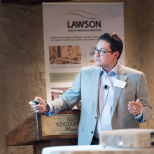
Dr. Sener maintains an active basic sciences and translational research laboratory focusing on gasotransmitter biology and therapeutics. Dr. Sener discussed the need to use “marginal” deceased donor kidneys - those from older donors, younger donors with existing medical issues, and donors post circulatory death – to treat end stage renal disease because due to a lack of donor supply.
Dr. Sener’s laboratory pioneered the use of hydrogen sulphide, a colourless gas with a strong “rotten egg” odor, to prolong organ storage, improve kidney re-perfusion, decrease dangerous inflammatory cells, promote quicker kidney function recovery, greater urine output and improve recipient survival.
Computer-assisted gastrointestinal surgery: Why can’t they see what I see?
Dr. Christopher Schlachta, Lawson scientist, medical director, Canadian Surgical Technologies and Advanced Robotics (CSTAR), LHSC
Image
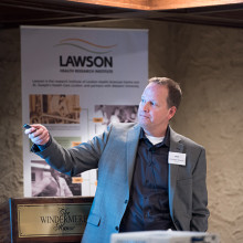
Dr. Schlachta’s current research interests are focused on development of computer-assisted surgical techniques and technologies to enhance care and training. Dr. Schlachta demonstrated how computer-assisted technologies in the operating room can enhance communication among surgeons and trainees to produce better outcomes for patients. He is currently partnering with industry to commercialize operating room technology he and a team of engineers at CSTAR have developed.
See more photos from this Café Scientifique on Lawson's Facebook page.
Cutting Edge: Surgical advancement through research
11:00pm - 1:00am
Before the bright lights of the operating room are turned on and the surgeons and operating room staff are gowned and ready, research conducted at Lawson Health Research Institute has backed many of the surgical innovations and firsts performed at London Health Sciences Centre and St. Joseph’s Health Care London. Join us to hear a panel of Lawson scientists discuss their cutting-edge work at this free community event.
In celebration of Canada’s 150th anniversary as a nation, this event is the second of a two-part series focusing on the future vision for health care in our country and the legacy our research will leave.
Date: Thursday, October 5, 2017
Time: 7 to 9 p.m. at Windermere Manor, 200 Collip Circle
Presented Talks
Computer-assisted gastrointestinal surgery
Dr. Christopher Schlachta
Transplant organ preservation: The best option may be “Stinky”
Dr. Alp Sener
Hand surgery: How small advances turn into complex surgical achievements
Dr. Bing Siang Gan
MODERATOR – Dr. David Hill
Registration
Registration is currently full. Please email @email to be placed on a wait list.
Event Information
Date: Thursday, October 5, 2017
Time: 7 to 9 p.m.
Location: Windermere Manor, 200 Collip Circle
… CARDIOVASCULAR INVESTIGATION UNIT REFERRAL FORM Cardiovascular Investigation Unit St. Joseph’s Hospital; B3-030 268 Grosvenor St. London, ON … CARDIOVASCULAR INVESTIGATION UNIT REFERRAL FORM Cardiovascular Investigation Unit St. Joseph’s Hospital; B3-030 268 Grosvenor …
CARDIOVASCULAR INVESTIGATION UNIT REFERRAL FORM Cardiovascular Investigation Unit St. Josephs Hospital; B3-030 268 Grosvenor St. London, ON N6A 4V2 Telephone: 519 646-6000 ext.64221 Fax: 519 646-629 PATIENT INFORMATION Surname: ___________________________ Given Name: ________________________________...
… CARDIOVASCULAR INVESTIGATION UNIT REFERRAL FORM Cardiovascular Investigation Unit St. Joseph’s Hospital; B3-030 268 Grosvenor St. London, ON … CARDIOVASCULAR INVESTIGATION UNIT REFERRAL FORM Cardiovascular Investigation Unit St. Joseph’s Hospital; B3-030 268 Grosvenor …
CARDIOVASCULAR INVESTIGATION UNIT REFERRAL FORM Cardiovascular Investigation Unit St. Josephs Hospital; B3-030 268 Grosvenor St. London, ON N6A 4V2 Telephone: 519 646-6000 ext.64221 Fax: 519 646-6292 PATIENT INFORMATION Surname: ___________________________ Given Name: _______________________________...
Cyclotron hits 10,000-bombardment milestone
Cyclotron staff at St. Joseph’s Health Care London have recorded a 10,000-mark milestone in the same understated way they work every day to improve patient care and cutting-edge research.
No balloons, no streamers, no fanfare: Just an efficient note atop a printout as the bombardment number spun past 9,999 in the early hours of Dec. 31.
“It’s taken us 15 years to get to this point and our work continues to grow,” says Michael Kovacs, PhD, Lead of Lawson’s Nordal Cyclotron & PET Radiochemistry Facility and Leader of the Imaging Research Program at Lawson Research Institute, the innovation arm of St. Joseph’s.
“The numbers are great but the real satisfaction is knowing every single bombardment means something important to a patient or a researcher working towards better patient health.”

St. Joseph’s GE PETtrace cyclotron is a particle accelerator that produces radioisotopes for use in positron emission tomography (PET) scans across Southwestern Ontario, from Windsor to Toronto. It is a vital tool for ultra-precise cancer diagnoses and for advanced research into scores of diseases.
In patient care, each “bombardment” – a grouping of radioisotopes that are then lab-processed, tested and made into smaller batches – can be used to aid cancer scans for as many as 25 people.
“A precise scan can make a dramatic difference, a life-changing difference, in how someone’s cancer is diagnosed and custom-managed,” Kovacs says. “If we think of the PET scanner as the engine of that transformative work, the cyclotron’s radioisotopes are its rocket fuel.”
Isotopes injected into patients are designed to have a short radioactive half-life – between two minutes and 110 minutes – which is another reason St. Joseph’s cyclotron is such an asset for timely care in the region.
“You can’t store or stockpile them. You have to use them almost immediately, so it’s essential to local and area hospital centres to have a ready, reliable source nearby,” Kovacs says.
About half the batched bombardments are used in patients to help with clinical diagnoses that will guide doctors’ treatment decisions.
The other half are used for research trials and pre-clinical research through Lawson, in fields as diverse as oncology, cardiology, neurology, psychiatry, metabolic disease and infectious diseases. In one promising study, for example, they’re being used to image specific brain proteins as researchers explore new disease-modifying treatment pathways for Alzheimer disease.
The next burgeoning field, Kovacs says, is theranostics: the science of diagnosing cancer and precision-attacking it at the same time. “That’s exciting for me, to be able simultaneously to see what we treat and treat what we see.”
About 15 highly specialized staff work at St. Joseph’s cyclotron facility, plus PhD-candidate researchers and other trainees.
Generous donors through St. Joseph’s Health Care Foundation have made much of this advanced research and next-level technology a reality. During the past few years, the Foundation granted nearly $800,000 in donor support to fund extensive renovations to the facility, making it possible to increase production of isotopes and expand life-saving care. Recently, $1 million in donations supported a new PET/CT scanner – the heart of Canada’s first national GE centre of excellence in molecular imaging and theranostics being developed at St. Joseph’s Hospital.
“We know the cyclotron is a critical tool in our imaging work and we are grateful to those donors who stepped up to help us with renovations that enabled the doubling of our facility’s production capability,” says Michelle Campbell, President and CEO of St. Joseph’s Health Care Foundation. “This renovation helps keep St. Joseph’s imaging program at the cutting edge of clinical care.”
The 40-tonne, room-sized cyclotron is more than a machine, and more than the experts who process, test, ship and use the radioisotopes, Kovacs notes.
It’s also testament to the vision of St. Joseph’s long-time chief medical physicist Frank Prato, PhD, and to the support of hospital administrators who saw its need and potential, he adds.
“We are innovators, and our vision is that we’re going to expand St. Joseph’s imaging expertise on an even larger world stage,” Kovacs says.
… is helping people get back behind the wheel who have lost their drivers’ license because of changes in their health … formerly a traffic helicopter pilot for a Toronto radio station, DARP gave him back his independence. He was taking … safe and accessible driving and those who need special instruction or vehicle adaptations to learn to drive. As well, …
Media Release July 22 2009 For Immediate Release Driver program helps grounded pilot regain independence London, Ontario - The driver assessment rehabilitation program (DARP) at Parkwood Hospital is helping people get back behind the wheel who have lost their drivers license because of changes in th...
Database funding could improve diagnosis and treatment of prostate cancer
A $125,000 grant from the Canadian Cancer Society will help create a database of PET/CT (positron emission tomography/computed tomography) and PET/MR (magnetic resonance) images of prostate cancer. The hope is that this database will be used to help improve diagnosis and treatment of men with prostate cancer.
The scans use radiopharmaceuticals to target prostate specific membrane antigen (PSMA), a transmembrane protein commonly found on prostate cancer cells.
“The idea behind this grant is to put together a database of PSMA PET/CT and PET/MR scans with annotated findings so medical professionals can scroll through cases and see the sites of prostate cancer. Our hope is this will help clinicians learn how to interpret these scans and ultimately help them to make informed treatment decisions for their patients,” says Dr. Katherine Zukotynski, Adjunct Scientist at Lawson Health Research Institute and lead researcher on the project.
The use of PSMA PET/CT and PET/MR scans in clinical practice is relatively new and currently only accessible through clinical trials. In fact, the first PSMA PET/MR scan in Canada was performed at St. Joseph’s Health Care London in 2016 by Dr. Glenn Bauman, Lawson Scientist and Radiation Oncologist at the London Regional Cancer Program at London Health Sciences Centre. Dr. Bauman is also part of this database project.
Studies have found these scans more accurately detect sites of prostate cancer than earlier imaging techniques, which then helps inform treatment decisions.
Dr. Zukotynski explains, “If you have an idea of the amount of disease and where it is located, and you can correlate it with prognosis, this could be very helpful. It might also allow physicians to compare current patients with patients who have similar findings, which may help determine the best therapies to try.”
There is hope that eventually this same database could lead to the use of artificial intelligence (AI) to assist in diagnosis and treatment planning.
“PSMA PET/CT and PET/MR may be tools helpful to categorize the total burden of disease, and then establish how that disease changes with therapy. Our first step down this path is to assemble a database that can be used both for research and educational purposes.”
The database will include data from centres across Canada, with a number of researchers contributing to the project funded by the Canadian Cancer Society.
Other principal investigators include: Dr. Bauman; Dr. François Bénard of the BC Cancer Research Institute; Dr. Vincent Gaudet, University of Waterloo; Dr. Phil Kuo, University of Arizona; Dr. Cynthia Ménard, Centre Hospitalier Universitaire de Montreal; and Dr. Ur Metser of the Princess Margaret Hospital. Dr. Carlos Uribe of the BC Cancer Research Centre and Dr. Aaron Ward of Lawson are co-applicants for the grant.
This is the part three of a three-part series on PSMA PET imaging research. Check out part one and two.
Research Area
Media Contact
Amanda Taccone
Communications Consultant & External Relations
Lawson Health Research Institute
T: 519-685-8500 ext. ext. 64059
C: 226-919-4748
@email
… Hill Integrated Vice President Lawson Health Research Institute Personal and Confidential Dear David: Office of the President 268 Grosvenor Street, PO Box 5777 London, ON N6A 4 V2 T: 519 646-6100 ext. … to you 1.5% of your annual base salary is based on St. Joseph's achieving the four targets outlined in our QIP. …
June 10, 2015 David Hill Integrated Vice President Lawson Health Research Institute Personal and Confidential Dear David: Office of the President 268 Grosvenor Street, PO Box 5777 London, ON N6A 4 V2 T: 519 646-6100 ext. 64200 F: 519 646-6054 www.sjhc.london.on.ca The purpose of this letter is to sh...
… Research and Scientific Director Lawson Health Research Institute Personal and Confidential Dear David: Office of the President 268 Grosvenor Street, PO Box 5777 London, ON N6A 4V2 T: 519 646-6100 ext. … to you, 1.5% of your annual base salary is based on St. Joseph's ach ieving the four indicators outlined in our …
June 4, 2018 David Hill Integrated Vice President Research and Scientific Director Lawson Health Research Institute Personal and Confidential Dear David: Office of the President 268 Grosvenor Street, PO Box 5777 London, ON N6A 4V2 T: 519 646-6100 ext. 64200 F: 519 646-6054 www.sjhc.london.on. ca The...
… base salary in the amount of $2,328.92. However, Bill 55, Strong Action for Ontario Act imposes limitations on the … to quality and continual improvement is evident. Lastly, I would also like to confirm the 2017/18 annual … performance based compensation goals are outlined in the St. Joseph's posted QIP. If you have any questions please do …
David Hill Page2 Confidential Given the corporate performance detailed above, you have achieved 66.67% of your performance-based compensation. This amounts to 1% of your annual base salary in the amount of $2,328.92. However, Bill 55, Strong Action for Ontario Act imposes limitations on the amount p...
… DEVELOPMENT FUND CALL FOR APPLICATIONS BACKGROUND Administered annually to nurses, or to support nursing development … Group or Team development ELIGIBILITY Courses or programs must be underway or completed in the current calendar year. Preference will be extended toward first-time applicants. APPLICATION PERIOD A call for …
DR. DAVID AND ZIVIA ANNE MELTZER NURSING PROFESSIONAL DEVELOPMENT FUND CALL FOR APPLICATIONS BACKGROUND Administered annually to nurses, or to support nursing development initiatives that contribute to the achievement of organizational and/or nursing priorities. Tuition associated with university o...