Search
Search
2373 Search Results:
… Health, (N) - Nurses, PSWs, (P) - Physicians, Pharmacists A minimum of 25% of this program is dedicated to … Convention Centre, London, Ontario 7:30 - 8:30 a.m. - REGISTRATION, EXHIBITORS & REFRESHMENTS 8:30 - 8:45 a.m. - … MD, Conference Chair, Division of Geriatric Medicine, St. Joseph’s Health Care London 8:45 - 9:45 a.m. - PLENARY …
LEGEND - WORKSHOP SUITED FOR: (A) - Allied Health, (N) - Nurses, PSWs, (P) - Physicians, Pharmacists A minimum of 25% of this program is dedicated to participant interaction. 33nd Annual Geriatric Medicine Refresher Day Current and Practical Knowledge to Inspire and Advance Care Wednesday, May 1, 20...
… in conjunction with the Geriatric Psychiatry programs at St. Joseph’s Health Care, London and London Health Sciences Centre, will host a conference today for health professionals focusing on … physician leader with the Geriatric Psychiatry Program at St. Joseph’s Regional Mental Health Care London, says: “The …
November 5, 2008 For Immediate Release International expert to address conference on complexity of mental illness in geriatric population London ON - The Division of Geriatric Psychiatry, in conjunction with the Geriatric Psychiatry programs at St. Josephs Health Care, London and London Health Scien...
… 1. Contact St. Joseph’s Health Care Foundation to advise us that you … this Gift of Securities Form. 2. If you work with an investment advisor, inform them of your plans to make a gift of … help you complete this form. 3. If you self-manage your investments, the Foundation is available to help you complete …
1. Contact St. Josephs Health Care Foundation to advise us that you wish to make a gift of securities and complete this Gift of Securities Form. 2. If you work with an investment advisor, inform them of your plans to make a gift of securities. They will help you complete this form. 3. If you self-ma...
Global initiative aims to prevent falls in older adults
Chaired by Dr. Manuel Montero-Odasso, Scientist at Lawson Health Research Institute, a group of 96 experts from 39 countries and 36 societies and agencies in Geriatric Medicine and Aging have come together to develop the “World Guidelines for Falls Prevention and Management for Older Adults: A Global Initiative.”
Published in Age and Ageing, the official journal of the British Geriatric Society, the guidelines provide recommendations to clinicians working with older adults to identify and assess fall risks.
“The global population is aging. Thanks to social and medical advances, some chronic conditions are diminishing proportionally. This is not the case for falls,” says Dr. Montero-Odasso, who is also a Geriatrician at St. Joseph’s Health Care London’s Parkwood Institute and a Professor at Western University’s Schulich School of Medicine & Dentistry. “Unfortunately, falls and related injuries among older adults are increasing and there is no sign of future decline.”
With new evidence and studies released since previous guidelines were published more than a decade ago, experts felt it was the right time for an update and an opportunity to incorporate a worldwide perspective
“Besides the rigorous methodology that 11 international working groups followed to provide new meta-analyses, including several new Cochrane collaborations, this World Falls Guidelines are, to the best of our knowledge, the first clinical practice guidelines in fall prevention to include a panel of older adults with lived experience in falls and mobility problems,” says Dr. Montero-Odasso. “They provided feedback, comments and opinions on our recommendations, making them considerably better and with wider applicability.”
Some key themes in the recommendations include:
- Falls can be prevented, but it requires multidisciplinary management.
- Preventing falls has wider benefits for quality of life.
- Fall risk can be assessed by trained clinicians with simple resources.
- A combination of interventions, including specific exercises to improve balance and strength, when delivered correctly, can effectively reduce fall risk in older adults.
Dr. Montero-Odasso says there was enough evidence to suggest that a global approach is needed to prevent falls in older adults and that “low risk does not mean no risk.” Even active older adults who are low risk should work on preventing loss of mobility and falls.
The next steps are for the guidelines to continue obtaining formalized endorsement of all the groups involved in the initiative and then for the guidelines to be fully implemented.
You can read the full guidelines here.
Scientist
Media Contact
Amanda Taccone
Communications Consultant & External Relations
Lawson Health Research Institute
T: 519-685-8500 ext. ext. 64059
C: 226-919-4748
@email
… The research institute of London Health Sciences Centre and St. Joseph’s Health Care London. LAWSON LAB GLOVE SWP … common surfaces and a gloved hand to carry the items. Best lab safety practice is to place the material in secondary … The research institute of London Health Sciences Centre and St. Joseph’s …
The research institute of London Health Sciences Centre and St. Josephs Health Care London. LAWSON LAB GLOVE SWP (SafeWorkPractice) (Wearing Gloves in Common Areas-Only when Required by Task) Gloves should NEVER touch door handles, elevator buttons, water fountains, telephones, card swipes, or any s...
… Thanks to donors who entrust us with their gifts, we are able to invest in critical health care needs in our community. 2014-2015 Grant Listing Over the year, St. Joseph’s Health Care Foundation … Thanks to donors who entrust us with their gifts, we are able to invest in critical …
Thanks to donors who entrust us with their gifts, we are able to invest in critical health care needs in our community. 2014-2015 Grant Listing Over the year, St. Josephs Health Care Foundation granted $4.9 million to St. Josephs Health Care London and Lawson Health Research Institute, the research ...
… 1 | P a g e 2015/16 LIST OF GRANTS PAID Patient Care Grants Hydrocollator, $2,955 - Mount Hope Centre for Long Term Care Physiotherapists will use a hydrocollator to provide therapeutic heat … Hope Centre for Long Term Care Fund and the Emerson C. Stewart Fund, items purchased for this initiative included …
1 | P a g e 2015/16 LIST OF GRANTS PAID Patient Care Grants Hydrocollator, $2,955 - Mount Hope Centre for Long Term Care Physiotherapists will use a hydrocollator to provide therapeutic heat that will help to treat residents for various conditions, including arthritis, soft tissue injury, musculoske...
… St. Joseph’s Health Care Foundation 2016-17 Allocations … $4,013,287 PATIENT CARE Chapel Refurbishment, $195,000 - St. Joseph's Hospital The 100-year-old chapel at St. Joseph’s Hospital received upgrades for a warmer and … St. Joseph’s Health Care Foundation 2016-17 Allocations …
St. Josephs Health Care Foundation 2016-17 Allocations MEDICAL COLLABORATION London Medical Innovation and Commercialization Network City Innovation Grant, $4,013,287 PATIENT CARE Chapel Refurbishment, $195,000 - St. Joseph's Hospital The 100-year-old chapel at St. Josephs Hospital received upgrades...
… St. Joseph’s Health Care Foundation 2017-18 Allocations … CARE Life Experiences in Complex Care, $3,049 – Parkwood Institute Main Building Patients in the Complex Care Program … & Care Needs at Parkwood Hospital, $7,100 – Parkwood Institute Main Building Medical and patient-related equipment …
St. Josephs Health Care Foundation 2017-18 Allocations PATIENT CARE Life Experiences in Complex Care, $3,049 Parkwood Institute Main Building Patients in the Complex Care Program were able to attend various community outings, while those who were unable to leave were provided with special on-site p...
… ST. JOSEPH’S HEALTH CARE FOUNDATION 2018-19 ALLOCATIONS … Wheelchair Inventory Refurbishment, $122,770 - Parkwood Institute Main Satellite Dialysis Program, $172,105 - Parkwood Institute Main Zero Suicide, $276,257 - Parkwood Institute … ST. JOSEPH’S HEALTH CARE FOUNDATION 2018-19 ALLOCATIONS …
ST. JOSEPHS HEALTH CARE FOUNDATION 2018-19 ALLOCATIONS PATIENT CARE Wheelchair Inventory Refurbishment, $122,770 - Parkwood Institute Main Satellite Dialysis Program, $172,105 - Parkwood Institute Main Zero Suicide, $276,257 - Parkwood Institute Mental Health Palliative Care Redevelopment, $941,619 ...
… Agency Sub-sector Public Hospital Organization Name St. Joseph's Health Care London Operation Name Operation … kWh 29,249 Cubic Meter 61,416.99485 35.98972 Parkwood Institute - Main Facilities used for hospital purposes 574,579 … 16,827 Metric Tonne 2,772,361.87125 40.91228 Parkwood Institute - RMHC Facilities used for hospital purposes 410,787 …
2018 Energy Consumption and Greenhouse Gas Emissions Reporting Sector Acute/Chronic Hospital Agency Sub-sector Public Hospital Organization Name St. Joseph's Health Care London Operation Name Operation Type Area Unit hrs/wk Quanity Unit Quanity Unit Quanity Unit Quanity Unit GHG Emissions (Kg) Energ...
… Agency Sub-sector Public Hospital Organization Name St. Joseph's Health Care London Operation Name Operation … kWh 25,435 Cubic Meter 54,253.06248 32.84142 Parkwood Institute - Main Facilities used for hospital purposes 574,579 … 18,328 Metric Tonne 3,010,701.21804 42.89784 Parkwood Institute - RMHC Facilities used for hospital purposes 410,787 …
2019 Energy Consumption and Greenhouse Gas Emissions Reporting Sector Acute/Chronic Hospital Agency Sub-sector Public Hospital Organization Name St. Joseph's Health Care London Operation Name Operation Type Area Unit hrs/wk Quanity Unit Quanity Unit Quanity Unit Quanity Unit GHG Emissions (Kg) Energ...
… N A W ◄ ► E y s ■ / / / / i~ SIJOSEPHs HEALTH CARE LONDON m t CJ 1:1 l!I m 4 2 1 Tim Hortons N $ N Ri ch m on d St re et W el lin gt on S tr ee t Grosvenor Street Cheapside Street Level 1 Entrance Level 1 Entrance … m t CJ 1:1 l!I m 4 2 1 Tim Hortons N $ N Ri ch m on d St re et W el lin gt on S tr ee t Grosvenor Street Cheapside …
N A W E y s / / / / i~ SIJOSEPHs HEALTH CARE LONDON m t CJ 1:1 l!I m 4 2 1 Tim Hortons N $ N Ri ch m on d St re et W el lin gt on S tr ee t Grosvenor Street Cheapside Street Level 1 Entrance Level 1 Entrance Level 0 Entrance Level 0 PARKING ELEVATORS TUNNEL ACCESS B A C D E F Grosvenor Parking Ga...
Groundbreaking Alzheimer’s and cancer studies receive $7.2M boost
Lawson Research Institute scientists and partners will focus on molecular imaging and theranostics to potentially transform the detection and treatment of neurodegeneration and cancer.
The quest to advance detection and treatment of Alzheimer’s disease and to personalize cancer care has received a major boost, with $7.2 million in funding to Lawson Research Institute (Lawson) of St. Joseph’s Health Care London (St. Joseph’s) for first-of-its kind research.
Lawson scientists will partner with a broad team of researchers at London Health Sciences Centre Research Institute (LHSCRI), McMaster University, University Health Network and BC Cancer on the ground-breaking studies focused on molecular imaging and theranostics as a potential game-changer in detecting and treating neurodegeneration and cancer, particularly prostate, brain and breast cancer.

Principal investigator Ting-Yim Lee, PhD, Lawson’s Director of PET/CT Research, and his team of investigators were awarded $2 million through the Ontario Research Fund – Research Excellence for the study titled “Improving Cancer and Alzheimer’s Disease Diagnosis and Treatment Through Cutting-edge Molecular Imaging and Theranostics”. Co-Principal Investigator is radiation oncologist Dr. Glenn Bauman at LHSCRI.
Additional funding from private-sector partners and Lawson, as well as from donors through St. Joseph’s Health Care Foundation, brings the total research investment to $7.2 million.
The research has the potential to offer hope for solutions to some of the most prevalent and pernicious diseases affecting Canadians, explains Lee.
“Both research projects are the first of their kind in Canada aimed at advancing how we diagnose and treat Alzheimer’s disease and cancer,” he says. “This collaborative funding initiative will also drive innovation in the exciting field of molecular imaging and theranostics at St. Joseph’s, at the heart of which is St. Joseph’s new, high-sensitivity GE HealthCare Omni Legend 2 PET/CT – the first in Canada.”
The studies encompass the following:
- Alzheimer’s disease: The new PET/CT at St. Joseph’s allows researchers to simultaneously study both blood flow and glucose metabolism in the brain. Both these mechanisms are believed to be contributing factors in the onset of Alzheimer’s. By measuring both at the same time, the research team hopes to uncover early signs that the brain is in trouble and at risk of plaque deposits and toxic proteins that have been linked to the development of Alzheimer’s.
- Cancer: The cancer study will focus on developing theranostic techniques to achieve personalized dosimetry – a method used to determine the exact amount of radiation a patient should receive during treatment, based on their individual characteristics. This maximizes effective treatment while minimizing harm to healthy tissues.
Molecular imaging and theranostics is a rapidly emerging field of medicine that combines ultra-precise scans and theranostics (a term that melds the words therapeutics and diagnostics). Together, they offer a one-two punch by integrating imaging and radiotracers that can identify the location and extent of diseased tissues and selectively destroy the abnormal cells while leaving surrounding healthy cells undamaged. In collaboration with GE HealthCare, St. Joseph’s is developing Canada’s first GE HealthCare Centre of Excellence in Molecular Imaging and Theranostics.
“By bridging the gap between research and clinical practice, we hope to ease the burden on patients and their families, offering more effective and compassionate care”
-Ting-Yim Lee, PhD, Director of PET/CT Research at Lawson Research Institute.
“We are already seeing the impact of novel theranostics for treatment of men with advanced prostate cancer,” says Bauman. “Promising new theranostic approaches are emerging for many cancers and this investment further positions London to be a leader in this area of research.”
In the initial phase of the studies, 100 patients will be recruited from St. Joseph’s Aging Brain and Memory Clinic at Parkwood Institute for the Alzheimer’s study; while 90 patients will be recruited from London Health Sciences Centre’s Verspeeten Family Cancer Centre for cancer studies. There are plans to recruit patients from the collaborating centres once results from the initial phase are confirmed.
“By bridging the gap between research and clinical practice, we hope to ease the burden on patients and their families, offering more effective and compassionate care,” says Lee. “We are deeply grateful for the opportunity to turn our research into real-world solutions that can make a meaningful impact.”
With dozens of 'firsts' in imaging research, “Lawson is a powerhouse of innovation,” adds Michael Kovacs, PhD, Program Lead, Lawson’s Imaging Research Program, and Lead, Cyclotron & PET Radiochemistry Facility. “We're excited to explore how this work could transform care."
Growing Tissues in the Lab
When challenged by surgeons to find better treatments for difficult-to-manage connective tissue diseases, Dr. David O’Gorman gladly accepted.
Dr. O’Gorman is a Molecular Biologist and Lawson Scientist based at St. Joseph’s Hospital, a part of St. Joseph’s Health Care London. His research focuses on understanding normal and abnormal connective tissue repair. He collaborates with researchers and clinicians working in many different disciplines, including those specializing in reconstructive surgery, orthopedics and urology.
Surgical reconstructions can be hampered by a lack of graft tissue, or graft tissue of insufficient quality, making it difficult to achieve optimal outcomes for the patients.
An example is a condition called urethral stricture disease (urethral scarring). This condition occurs in males and typically causes symptoms such as frequent and urgent urination, and slow urinary stream. In extreme cases, it can cause urinary tract infections, permanent bladder dysfunction and renal failure. Recurrence rates after minimally invasive treatments are high, and so many urologists recommend open surgical approaches.
Surgeons can use the patient’s own tissues to reconstruct the urethra after stricture removal. This tissue is normally sourced from the buccal cavity in the mouth but taking large tissue grafts can result in complications. In cases where buccal grafts have been used for previous reconstructions, there may not be enough intact tissue left.
Dr. O’Gorman sees a solution in growing sheets of human buccal tissues in the lab.

“We are currently using buccal graft trimmings as a source of cells, culturing them in a 3D environment and expanding them to create tissues of suitable size, density and elasticity.”
The patient’s own cells are used to generate a tissue graft for urethral reconstruction. While several research groups have developed this approach in the past, few have attempted to translate their models for clinical use.
“Our immediate goal is to provide proof of principle – that we can consistently generate grafts of suitable size and functional characteristics,” explains Dr. O’Gorman, “In the future, we could be providing bioengineered graft tissues for reconstructive surgeries here in London.”
Bioengineered human tissues can also be used as ‘mimetics’ – replications of human tissues – to study diseases, especially those difficult to model using routine laboratory methods.
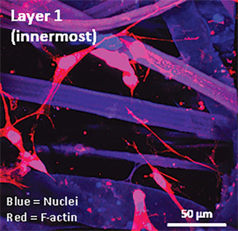
Instead of a using a growth media or sterile plastic dishes, 3D cell culture is achieved by embedding cells in a matrix of proteins and other molecules normally found in those tissues. In this environment, gene expression and growth is more similar to cells of connective tissues in the body being replicated.
Dupuytren’s disease (or Dupuytren’s Contracture) affects the palmar fascia in the hand, a connective tissue beneath the skin that extends from the base of the palm into the fingers. This disease can be understood as a type of excessive scarring, where normal tissue repair processes have gone awry and dense scar tissue forms, typically causing permanent palm or finger flexion that restricts hand function.
This condition is surprisingly common and may affect more than one million people in Canada. While there are surgical treatment options available, none consistently prevent this disease from recurring in at least a third of patients.
“Due to its high recurrence rate after treatment, Dupuytren’s disease is currently considered incurable. Our challenge is to understand it well enough to develop truly effective treatments,” says Dr. O’Gorman.
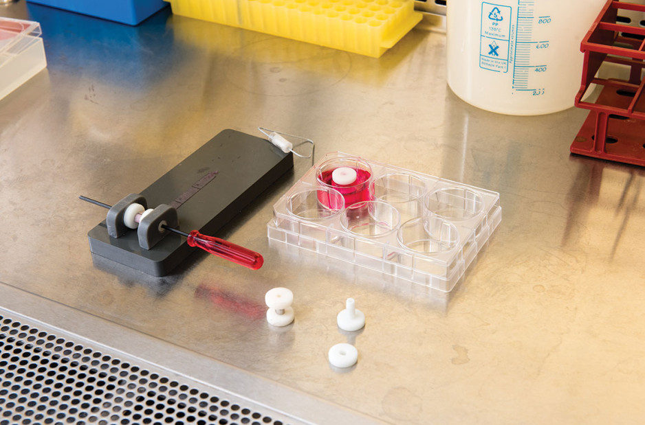
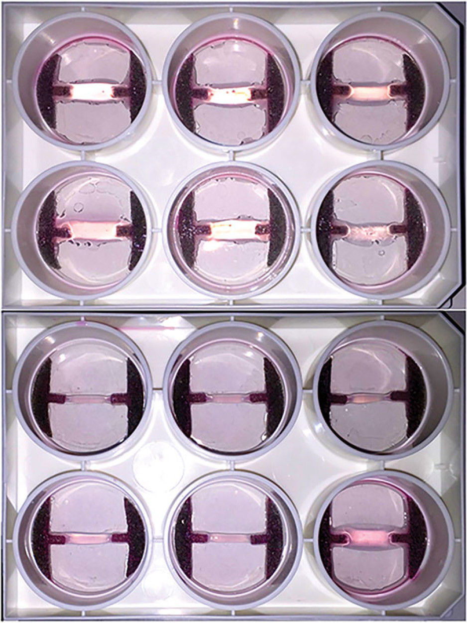
Human hands have unique characteristics not found in other species, making animal models impractical. Instead, Dr. O’Gorman’s team extracts cells from the diseased palmar fascia of patients undergoing hand surgeries and bioengineers them into palmar fascia ‘contractures’ in the lab.
“Since the cells from a single palmar fascia sample can be used to grow dozens of little contractures, we can test many different treatments simultaneously to see what works best for each patient.”
This approach may also allow them to determine if Dupuytren’s disease is truly one disease, or a group of similar diseases that cause palm and finger contractures.
“Often, Dupuytren’s disease is clearly heritable, but some individuals have no family history of it and develop apparently sporadic disease,” notes Dr. O’Gorman. “We want to determine if these are truly the same disease at the molecular level.”
Another major cause of abnormal connective tissue repair is infection, and tissue mimetics can play a role here, too. While rare, infections of artificial joint replacements are particularly devastating for patients, as they typically require readmission to hospital to remove the infected joint, weeks of antibiotic-based treatment, and an additional surgery to replace the artificial joint.
In addition to the associated pain and suffering, these procedures are technically challenging and costly to our health care system.
Artificial shoulder joint infections are most frequently caused by the microorganism Cutibacterium acnes (C. acnes). C. acnes infections disrupt normal tissue repair processes after surgery, cause shoulder tissues to die and promote loosening of the artificial joint. These infections are difficult to diagnose, and there is a lack of reproducible
models in which to study them. Dr O’Gorman’s team has set out to create the first human Shoulder-Joint Implant Mimetic (S-JIM) of C. acnes infection.
“While S-JIMs are more complex, they are 3D in vitro cell culture systems designed to mimic human tissues, like those that we use for studying Dupuytren’s disease.”
S-JIMs include layers of artificial human tissue, wrapped around cores of titanium alloy or cobalt chrome, the metals used to create artificial joints. They are co-cultured with C. acnes under low oxygen conditions similar to those that normally occur around artificial shoulder joints.
“We are bioengineering simple 3D cell cultures to more closely mimic the complexity of human tissues, with blood supply, nerves and interactions with other cells.” – Dr. David O’Gorman
Studying the connective tissue layers close to the infection allows researchers to investigate processes that promote infection, such as the formation of a biofilm that harbours and protects the bacteria from the body’s immune system. They are also able to test whether novel treatments can disrupt biofilm formation and increase the effectiveness of antibiotics.
Dr. O’Gorman predicts that in the future, medical researchers will routinely use bioengineered 3D human tissue and organ mimetics to accelerate our understanding of disease.
“The technology is in its infancy, but the potential for using bioengineered human tissues for surgical reconstructions or as disease models is huge. At Lawson, we’re ready to take on health care challenges and build on innovative approaches to improve the quality of life for patients.”
ONLINE EXCLUSIVE: What is 3D cell culture?
Medical researchers have grown human cells in culture media on or in sterile plastic dishes, such as Petri dishes, for more than 50 years.
Some cells, such as blood cells, can survive and grow in suspension, while others like smooth muscle cells need¬ to adhere to a surface to survive and grow. These are often called “2D cell cultures” because the cells grow horizontally across the bottom of the dish.
Some cells derived from connective tissues, such as fibroblasts, are not only adherent, but also very sensitive to the stiffness of their environment (“biomechanically sensitive” cells). Plastic dishes are at least 10,000 times stiffer than most connective tissues, and when biomechanically sensitive cells detect stiff surfaces, they can change the expression of their genes and behave abnormally.
The most common proteins in these tissues - and in the entire human body - are collagens, and one routine 3D cell culture approach is to embed fibroblasts in a collagen gel (gelatin). Fibroblasts in this environment can grow in any direction they choose, and their gene expression is more similar to cells in connective tissues.
These simple 3D cell cultures represent tissue engineering in its most basic form.
“Our challenge is to bioengineer simple 3D cell cultures in the lab to more closely mimic the complexity of human tissues, which have blood supply, nerves and interactions with other cells and tissues that modify their function and ability to heal after injury,” explains Dr. O’Gorman.
Dr. David O’Gorman is a Lawson Scientist and Co-director, Cell and Molecular Biology Laboratory at The Roth | McFarlane Hand and Upper Limb Centre in London, Ontario. He is also an Assistant Professor at Western University.
… The team at the Diabetes Education Centre of St. Joseph’s Health Care London would like to thank … diabetes support for individuals with diabetes in southwestern Ontario. We would also like to extend our appreciation … 1-1 Pump Start Information for Patients … 1.1 2. Before Starting on a Pump …
Resource Guide to Insulin Pump Success Acknowledgements The team at the Diabetes Education Centre of St. Josephs Health Care London would like to thank Sanofi-Aventis for their generous contribution to sponsoring this educational initiative, thereby encouraging further development of resources and d...
… Student Contract Health Requirements_20160817 Page 1 of 2 GUIDELINE TO HEALTH REQUIREMENTS FOR STUDENT PLACEMENT INTRODUCTION All post-secondary students obtaining learning experience and all … Student Contract Health Requirements_20160817 Page 1 of 2 GUIDELINE TO HEALTH REQUIREMENTS FOR STUDENT PLACEMENT INTRODUCTION All post-secondary students …
Student Contract Health Requirements_20160817 Page 1 of 2 GUIDELINE TO HEALTH REQUIREMENTS FOR STUDENT PLACEMENT INTRODUCTION All post-secondary students obtaining learning experience and all faculty members working with students shall comply with the health and safety requirements for staff, as spe...
… MIND & SPIRIT SINCE 1869 Renowned for compassionate care, St. Joseph’s is one of the best academic health care organizations in Canada dedicated to helping people live to their fullest by minimizing the effects of injury, disease and … MIND & SPIRIT SINCE 1869 Renowned for compassionate care, St. Joseph’s is one of the best academic health care …
CARING FOR THE BODY, MIND & SPIRIT SINCE 1869 Renowned for compassionate care, St. Josephs is one of the best academic health care organizations in Canada dedicated to helping people live to their fullest by minimizing the effects of injury, disease and disability through excellence in care, teachin...
… ' ~ i S! JOSEPHs HEALTH CAR[ LON DON Policy SJ_GEN053 Essential … ongoing need to protect long-term care home residents and staff from the risk of COVID-19, particularly as residents … the mental and emotional well-being of residents and strive to be as accommodating as possible when scheduling …
' ~ i S! JOSEPHs HEALTH CAR[ LON DON Policy SJ_GEN053 Essential Caregiver Visitor Policy for Mount Hope Appendix B Guiding Principles Under the Fixing Long-Term Care Act, 2021, residents have a right to receive visitors. At a minimum, residents are permitted to have four indoor visitors (combination...