CIBC donation allows scientists to visualize the possibilities in cancer care
The confocal microscope gives medical researchers the ability to observe physiological processes unfolding inside cells and tissue in real time
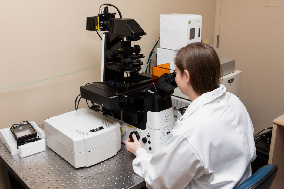
A generous donation from CIBC is giving scientists at Lawson Health Research Institute an impressive new tool to study illness and disease. The $240,000 gift to St. Joseph’s Health Care Foundation was used to purchase a confocal microscope, state-of-the-art technology that is supporting medical researchers in the quest to find and test new treatments for diseases like Alzheimer’s, diabetes, kidney stones and breast cancer.
“This gift is a great tribute to medical researchers and the meticulous work they continuously do to improve patient care for people in our region,” says Michelle Campbell, President and CEO, St. Joseph’s Health Care Foundation.
Today’s announcement falls on World Cancer Day, a global initiative to raise awareness of cancer and advocate for prevention, detection and treatment. Inspired by its ambition to help change the future of cancer, CIBC is continually investing in innovative cancer research.
“We’re proud to support the talented team at St. Joseph’s in their mission to create a healthier future for all with this donation,” says Jonathon Dent, Senior Vice-President, Region Head, Ontario West at CIBC. “Their courageous work and focus on innovation will bring us one step closer to realizing our collective ambition of creating a future without cancer.”
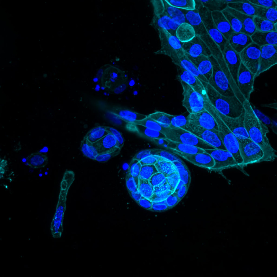
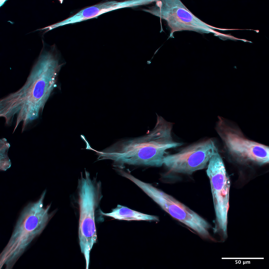
Using the confocal microscope, scientists can see the cellular world in vivid, three-dimensional images lit up in fluorescent colours. The images enable scientists to observe physiological processes unfolding inside cells and tissues in real time. This insight gives scientists the ability to compare normal and abnormal tissue samples in the laboratory.
“Tools like the confocal microscope act as a bridge between our clinics and the research lab,” says Dr. Jeremy Burton, Chair in Human Microbiome and Probiotics. “It enables scientists and physicians to collaborate on new therapies and develop a better understanding of disease.”
Lawson was the first research institute in Canada to install this advanced and important instrument. Today, it is one of four in the region being used to observe and study disease development and progression.
Dr. Burton recently collaborated with Dr. Muriel Brackstone, surgical oncologist and Medical Director of St. Joseph’s Breast Care Program, on a study to assess whether it is possible to change the bacterial microbiome in the breast with probiotics. They relied on the confocal microscope to observe the internalization between probiotic bacteria and special types of immune cells. Their findings, which will be published in an upcoming research paper, could reveal new insights about the future of breast cancer treatment.
"I am so grateful for CIBC’s donation, which has given scientists at St. Joseph’s access to first-rate medical research equipment to conduct clinical trials that answer important scientific questions and ultimately improve patient outcomes,” says Dr. Brackstone.