Your donation matters to Stephanie
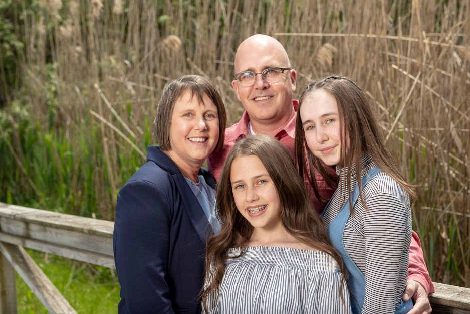
When Stephanie Wilds was 45 she was diagnosed with invasive breast cancer in both breasts. A mom to two young girls, she feared the worst: that she wouldn’t make it past five years.
Stephanie underwent a lumpectomy performed by Dr. Muriel Brackstone at St. Joseph’s Breast Care Program, along with 15 months of chemotherapy, immunotherapy and six weeks of radiation therapy. Three years later Stephanie is still cancer-free.
During a lumpectomy, surgeons remove the tumour and a small amount of normal tissue around it to ensure the cancer doesn’t return. Determining the exact spot where the cancer ends and healthy tissue begins is challenging using standard imaging equipment. For patients, this means they may have to return for additional surgery to catch it all.
Because of your support, Dr. Jeffrey Carson, a medical researcher at Lawson Health Research Institute, is perfecting intraoperative photoacoustic tomography to advance lumpectomies. This imaging tool uses light absorption to show the difference between healthy and cancerous tissue.
Dr. Carson, together with Dr. Brackstone and radiologist Dr. Anat Kornecki, are able to scan surgically removed samples to determine more accurately and almost immediately whether all of the cancer has been removed. This means people like Stephanie have peace of mind when returning home to her family.
When I was diagnosed with invasive breast cancer I didn’t have hope that I would be here in five years.