Search
Search
3187 Search Results:
Dr. s copy ( Western Schulich School of Medicine & Dentistrys copy ( Department of s copy ( Medical Affairs copy ( THIS LETTER OF OFFER IS ONLY TO BE ISSUED AFTER CONFIRMATION OF THREE SATISFACTORY REFERENCES FOR THE CANDIDATE. Dea...
Dr. s copy ( Western Schulich School of Medicine & Dentistrys copy ( Department of s copy ( Medical Affairs copy ( THIS LETTER OF OFFER IS ONLY TO BE ISSUED AFTER CONFIRMATION OF THREE SATISFACTORY REFERENCES FOR THE CANDIDATE. Dea...
How changes in the brain affect walking while talking in older adults
Dr. Manuel Montero-Odasso’s research demonstrates that gait testing, such as walking while performing a cognitively demanding task like counting backwards (dual-task gait),can be an effective predictor of progression to dementia. In a new study, a team at Lawson Health Research Institute and Western University’s Schulich School of Medicine & Dentistry has discovered changes to the brain that correspond to these findings.
These changes identify a brain mechanism that corresponds with slow dual-task gait among older adults with mild cognitive impairment (MCI), an intermediate stage between the expected cognitive decline of normal aging and the more serious decline of dementia. Through their work Dr. Montero-Odasso and his team have found that a high dual-task gait cost, or a significant slowdown in walking speed when dual-tasking, is associated with a two- to three-fold increased risk of progression to dementia. However, the brain mechanism underlying this association was unclear.
To address this research question, Dr. Montero-Odasso partnered with Robert Bartha, PhD, an imaging scientist at Schulich Medicine & Dentistry and Robarts Research Institute at Western University. The team used magnetic resonance imaging (MRI) to examine the medial temporal areas of the brain, particularly the hippocampus, the parahippocampal gyrus, and the entorhinal cortex, which are regions particularly vulnerable to degeneration in Alzheimer’s disease. Participants were 40 older adults with MCI taking part in Dr. Montero-Odasso’s “Gait and Brain Study” at St. Joseph’s Health Care London’s Parkwood Institute.
The researchers found that participants with higher dual-task gait costs had a smaller grey matter volume in the left entorhinal cortex. Although grey matter volume loss is a common finding in people with Alzheimer’s disease, it is still unclear which areas of the brain are first affected by neurodegeneration. This finding points to the entorhinal cortex as a susceptible brain region in early stages of cognitive decline. This is in line with previous studies reporting that progression to Alzheimer’s disease is associated with volume loss in the entorhinal cortex.
The study therefore suggests that cognitive and motor dysfunction in older adults with MCI share common changes to the brain. This further supports that dual-task gait changes may be a measurable motor marker for neurological degeneration happening in Alzheimer’s disease.
“These novel results show that early brain changes common to pre-dementia states can be manifested by the way patients walk,” says Dr. Montero-Odasso, scientist at Lawson, geriatrician at St. Joseph’s Health Care London, and professor at Schulich Medicine & Dentistry. “This evidence supports walking while performing a cognitively demanding task as an important way to help predict dementia.”
The study, “Entorhinal Cortex Volume Is Associated With Dual-Task Gait Cost Among Older Adults With MCI: Results From the Gait and Brain Study,” is published in The Journals of Gerontology: Series A.
Scientist
… FOR THE BODY, MIND & SPIRIT SINCE 1869 \~ SIJOSEPHs HEALTH CARE LONDON Evacusled Deployment Evacusleds are used to … FOR THE BODY, MIND & SPIRIT SINCE 1869 \~ SIJOSEPHs HEALTH CARE LONDON Evacusled Preparedness – Step 1 Clear the your … FOR THE BODY, MIND & SPIRIT SINCE 1869 \~ SIJOSEPHs HEALTH CARE LONDON Evacusled Preparedness – Step 2 Prepare the bed …
CARING FOR THE BODY, MIND & SPIRIT SINCE 1869 \~ SIJOSEPHs HEALTH CARE LONDON Evacusled Deployment Evacusleds are used to assist with the evacuation of nonambulatory residents and patients. At Mount Hope and Parkwood Institute Main Building...
… Critieria for admission to LTV Program Feb 2024.doc Complex Care Program – Parkwood Institute Patients Eligible for … Term Ventilation Beds There are six beds within the Complex Care Program at Parkwood Institute that provide a clinical … purpose of transferring these individuals to the Complex Care Program is to provide a more appropriate setting to …
Critieria for admission to LTV Program Feb 2024.doc Complex Care Program Parkwood Institute Patients Eligible for transfer to the Long Term Ventilation Beds There are six beds within the Complex Care Program at Parkwood Institute that provide a clinical setting for individuals requiring long-term m...
… How to refer a patient to Palliative Care Palliative Care provides pain management and relief of symptoms to … progressive or terminal illness. Palliative Care focuses on physical, psychological, social and … How to refer a patient to Palliative Care Palliative Care provides pain management and relief of …
How to refer a patient to Palliative Care Palliative Care provides pain management and relief of symptoms to those experiencing life-threatening, progressive or terminal illness. Palliative Care focuses on physical, psychological, social and spiritual needs while remaining sensitive to the uniquenes...
… INTEGRATION NETWORK (the “LHIN”) AND St. Joseph's Health Care, London (the “Hospital”) WHEREAS the LHIN and the … Michael Barrett, CEO Date St. Joseph's Health Care, London By: _________________________________ … Bilateral Hip/Knee Replacement 714 St. Joseph's Health Care London St. Joseph's Health Care London NON-LHIN FUNDING …
H-SAA Amending Agreement Extension to March 31, 2017 Page 1 H-SAA AMENDING AGREEMENT THIS AMENDING AGREEMENT (the Agreement) is made as of the 1st day of October, 2016 B E T W E E N: SOUTH WEST LOCAL HEALTH INTEGRATION NETWORK (the LHIN) AND St. Joseph's Health Care, London (the Hospital) WHEREAS t...
… intake of foods • Increase diabetes medication on advice of care provider Prevent By • Consistency in adhering to meal … intake of foods • Increase diabetes medication on advice of care provider Prevent By • Consistency in adhering to meal …
Guidelines for Managing Hyperglycemia (High Blood Glucose) Signs and Symptoms of Hyperglycemia ( High Blood Glucose ) ONSET Gradual (hours to days) USUAL CAUSES Illness, infection, surgery, injury Stress: emotional or physical Too little insulin Increased food Exercise (in type 1) with blood g...
… intake of foods • Increase diabetes medication on advice of care provider Prevent By • Consistency in adhering to meal … intake of foods • Increase diabetes medication on advice of care provider Prevent By • Consistency in adhering to meal …
Guidelines for Managing Hyperglycemia (High Glucose Level) Signs and Symptoms of Hyperglycemia ( High Glucose Level ) ONSET Gradual (hours to days) USUAL CAUSES Illness, infection, surgery, injury Stress: emotional or physical Too little insulin Increased food Exercise (in type 1) with glucose...
… better symptom improvement than using iCBT alone. As remote care, such as iCBT, has more then doubled since the … This research was supported by the St Joseph’s Healthcare Foundation Is iCBT for adult military populations an … better symptom improvement than using iCBT alone. As remote care, such as iCBT, has more then doubled since the …
Internet-Based Cognitive Therapy (iCBT) is the delivery of cognitive behavioral therapy (CBT) through a computer, phone, or mobile device, often guided by a mental health professional. Veteran & active military members are unique populations for treatment. They have distinct symptom presentations & ...
ICES launches call for applications to its next faculty training cycle
The ICES Faculty Scholars Program has officially launched the call for applications to its fourth training cycle (2019-2021).
This two-year, part-time learning opportunity is open to Ontario-based academic researchers who are passionate about population health and health services research, and wish to obtain an appointment as an ICES Scientist,
The program was established at ICES Western in 2013 as part of its strong focus on supporting trainees and new investigators. By leveraging partnerships with other ICES sites and in conjunction with an interactive e-learning environment, faculty from different regions are able to fully participate in the ICES Faculty Scholars program.
Scholars will have access to:
- Training in the use of Ontario’s health administrative databases for research.
- Seminars on advanced methods relevant to population health research.
- Opportunities for collaboration with experts across the ICES network.
- Individual project mentorship and data analytic support to develop and complete one or more research studies using Ontario’s health administrative databases.
Hear from successful ICES Faculty Scholars:
“The methods of planning and completing an ICES project are unique. This program is the best possible way to demystify the process and produce high quality work from the outset. The amount of support is outstanding, so you have confidence that your work will be successful.”
Dr. Andrew Appleton, Assistant Professor, General Internal Medicine, Western University
“The Faculty Scholars Program is extremely well structured and run. The Program provides a positive and open learning environment for Scholars to ask questions and learn about the administrative and practical aspects pertaining to conducting research at ICES.”
Dr. James Crispo, Researcher, Health Sciences North Research Institute
Applications for the 2019–2021 program will be accepted until January 18, 2019. Any questions regarding the program, applications or eligibility can be directed to @email.
Go to the ICES website for more information on the ICES Faculty Scholars Program.
ICES is a not-for-profit research institute encompassing a community of research, data and clinical experts. With its growing network across the province, ICES researchers are able to access a vast and secure array of health-related data to evaluate health care delivery and outcomes. Learn more about ICES Western.
ICU patients with non-brain-related injuries may suffer undetected cognitive dysfunction
LONDON, ON - A new study led by Western University and Lawson Health Research Institute has found that most patients entering hospital intensive care units (ICU) for non-brain-related injuries or ailments also suffer from some level of related cognitive dysfunction that currently goes undetected in most cases.
The findings were published today in the influential scientific journal, PLOS One.
Many patients spend time in the ICU for reasons that have nothing to do with a known brain injury, and most health care providers and caregivers don’t have any evidence to believe there is an issue with the brain. For example, a patient may have had a traumatic injury that does not involve the brain, yet still requires breathing support to enable surgeons to fix damaged organs, they may have issues with their heart or lungs, they may contract a serious infection, or they may simply be recovering from a surgical procedure like an organ transplant that has nothing directly to do with their brain.
For the study, Western researchers from the Schulich School of Medicine & Dentistry and the Brain and Mind Institute and researchers from Lawson assessed 20 such patients as they left the ICU and every single patient had detectible cognitive deficits in two or more cognitive areas of investigation, including memory, attention, decision-making and reasoning. Again, this is in spite of the fact that, on the face of it, they had no clear brain injury.
The discovery was made using online tests, developed by renowned Western neuroscientist Adrian Owen and his teams at the Brain and Mind Institute and BrainsCAN, which were originally designed to examine cognitive ability in patients following brain injuries but for this purpose, are being used to detect cognitive deficits in people who have spent time in an intensive care unit without a diagnosed brain injury.
“Many people spend time in an intensive care unit following a brain injury and, of course, they often experience deficits in memory, attention, decision-making and other cognitive functions as a result,” explains Owen, a professor at Schulich Medicine & Dentistry. “In this study, we were interested to see how patients without a specific brain injury fair after leaving the ICU. The results were astonishing.”
Why cognitive ability declines even in non-brain related visits to the ICU likely varies from patient to patient, but Dr. Kimia Honarmand from Schulich Medicine & Dentistry says the lesson to be learned is that many conditions affect brain function, even though they might not directly involve the brain.
“If you are having trouble breathing, your brain may be starved of oxygen. If you have a serious infection, the inflammation that occurs as a result of infection may affect brain function. If you are undergoing major surgery, you might be given drugs and have procedures that may affect your breathing, which in turn may affect the flow of oxygen to the brain,” explains Dr. Honarmand. “What we have shown here is that all or any of these events can lead to deficits in brain function that manifest as impairments in cognition. And healthy cognition is a vital determinant of functional recovery.”
Dr. Marat Slessarev, Lawson Scientist, says these findings can shift how the medical community treats incoming patients and more importantly, outpatients following ICU visits.
“Historically, the clinical focus has been on just survival. But now we can begin to focus on good survival,” says Dr. Slessarev, also an associate member at the Brain and Mind Institute and an assistant professor at Schulich Medicine & Dentistry. “These sensitive tests will enable doctors to both detect cognitive impairment and track cognitive performance over time, which is the first step in developing processes for optimizing brain recovery.”
-30-
About Western
Western delivers an academic experience second to none. Since 1878, The Western Experience has combined academic excellence with life-long opportunities for intellectual, social and cultural growth in order to better serve our communities. Our research excellence expands knowledge and drives discovery with real-world application. Western attracts individuals with a broad worldview, seeking to study, influence and lead in the international community.
About The Schulich School of Medicine & Dentistry
The Schulich School of Medicine & Dentistry at Western University is one of Canada’s preeminent medical and dental schools. Established in 1881, it was one of the founding schools of Western University and is known for being the birthplace of family medicine in Canada. For more than 130 years, the School has demonstrated a commitment to academic excellence and a passion for scientific discovery.
About Lawson Health Research Institute
Lawson Health Research Institute is one of Canada’s top hospital-based research institutes, tackling the most pressing challenges in health care. As the research institute of London Health Sciences Centre and St. Joseph’s Health Care London, our innovation happens where care is delivered. Lawson research teams are at the leading-edge of science with the goal of improving health and the delivery of care for patients. Working in partnership with Western University, our researchers are encouraged to pursue their curiosity, collaborate often and share their discoveries widely. Research conducted through Lawson makes a difference in the lives of patients, families and communities around the world. To learn more, visit www.lawsonresearch.ca.
Media Contacts
Celine Zadorsky
Senior Media Relations Consultant
Communications & Public Engagement
T: 519-685-8500 ext. 73502
Celine.zadorsky@lhsc.on.ca
Illuminating the body's smallest secrets
Lawson Research Institute’s cyclotron facility is a formidable partner in the fight against disease.
Tucked behind the protective lower walls of St. Joseph’s Hospital lies a futuristic workshop of sorts – a place where science, technology and leading-edge medical care converge.
Within a behemoth, 62-ton machine, a swirling vortex of powerful magnetic fields and electric pulses is creating bursts of radioactive isotopes – tiny, potent sparks of life-saving potential. In the hands of technologists, researchers and clinicians, these chemical elements become diagnostic tracers and therapeutic agents, each particle revealing secrets of the human body.
The machine is a cyclotron - a type of particle accelerator and the only one of its kind in the region. At Lawson Research Institute’s Cyclotron & PET Radiochemistry Facility, scientists are producing a steady and timely supply of short-lived radioisotopes every day to study, detect and treat disease.

These radioisotopes become a beacon in positron emission tomography (PET) scans, illuminating the hidden shadows of cancer and other diseases. Others provide a precise map of the intricate pathways of blood flow, biological functions, location of specific cells and proteins, and the body’s skeletal architecture.
A formidable partner in the fight against disease, “the cyclotron facility is a hub for Southwestern Ontario that is uncovering the possibilities for improving patient care in numerous ways,” says Michael Kovacs, PhD, Lead, Lawson’s Cyclotron & PET Radiochemistry Facility and Program Lead, Lawson Imaging Research Program.
St. Joseph’s cyclotron supports a wide variety of research projects including imaging applied to oncology, cardiology, neurology, psychiatry, metabolic disease, infectious diseases, bioelectromagnetics and other areas.
“The scope of discoveries already making a difference, and the possibilities within reach, are a source of great pride for Lawson and for London,” adds Frank Prato, PhD Lawson scientist and Chief Medical Physicist at St. Joseph’s.
For Kovacs, Prato, their teams and partners, St. Joseph’s cyclotron is a testament to ingenuity and innovation, a world where every spin and burst of charged particles brings a promise of hope and healing.
Powering innovation
Generous donors to St. Joseph’s Health Care London have made both advanced research and next-level technology a reality. During the past few years, more than $1.1 million in donations funded extensive renovations to the Cyclotron & PET Radiochemistry Facility, making it possible to increase production of isotopes and expand life-saving care.
Recently, $1 million in donations supported a new PET/CT – the heart of the Canada’s first national GE centre of excellence in molecular imaging and theranostics being developed at St. Joseph’s Hospital.
Imaging
Lawson is in the international forefront of medical imaging
Research that takes a deep look inside the human body and seeks to understand how tissues, bones, organs and cells work, and then guides next steps when the body doesn’t work as it should.
At Lawson, we see things more clearly than anyone.
Lawson leads medical and imaging research. Learn more:
We’re home to a host of Canadian and world firsts that have resulted in new pathways of diagnosis, treatment and care.
We’re recognized as pioneers in molecular imaging: a virtual alphabet of MRIs, PET scans, CT scans and more.
With technologies that also include our state-of-the-art cyclotron, we keep pushing the boundaries of these technologies so that soon we hope to test treatments with the same pinpoint precision and technology we use for imaging. This is a new and exciting field called theranostics, and it offers the very real possibility of a magic bullet to eradicate some cancers.
Our award-winning scientists, trainees and staff share their discoveries to improve patient lives locally and around the world.
Lawson is a global leader in imaging research. Read on to see where it can lead you.
Lawson Intranet (password protected) | ICES: data-focused research | Animal Care Committee
Imaging “hidden” regions of the heart
After suffering a heart attack, some patients develop a microvascular obstruction, an area of the injured heart with extremely poor blood flow. These patients are at an increased risk of developing heart failure in the future.
Medical imaging technologies such as magnetic resonance imaging (MRI) and positron emission tomography (PET) can be used to study the remodeling process after a heart attack that can lead to a microvascular obstruction. However, poor blood flow makes it difficult to get contrast agents into the obstruction. Contrast agents are used in medical imaging to show contrast between different types of tissue, such as damaged and healthy tissue.
Benjamin Wilk, a PhD candidate at Lawson Health Research Institute and Western University’s Schulich School of Medicine & Dentistry, will investigate whether a hybrid PET/MRI system and a new method of administering contrast agents can allow researchers to image microvascular obstructions and study these “hidden” regions in the heart.
Contrast agents are usually injected as a bolus, meaning the entire injection is administered immediately. In this study, participants will instead receive a constant infusion of an MRI contrast agent and PET tracer, which means the injection will be delivered over the course of an hour. The MRI contrast agent they are using is sensitive to blood flow and scar tissue, and the PET tracer is sensitive to inflammatory cells.
This will allow researchers to study the anatomy, blood flow and inflammatory processes in microvascular obstructions a week after heart attack. Participants will then be imaged again after six weeks to study the long-term effects on heart function.
“Studying the heart after a heart attack using novel contrast agent injection strategies with simultaneous PET/MRI could provide crucial information for treatment planning, helping us reduce the number of people affected by heart failure in the future,” says Wilk. “This project could also lead to further research into finding better ways to administer PET tracers and MRI contrast agents. These methods could be applied to different diseases as well.”
Wilk received a Lawson Internal Research Fund (IRF) Studentship to conduct the study, which will be supervised by Dr. Frank Prato, Assistant Director, Lawson and leader of the Lawson Imaging research program at St. Joseph’s Health Care London.
“Lawson's IRF is valuable for students for many reasons. It not only allows us to conduct further research, it also enriches our experience by giving us opportunities to write grants and attend conferences,” adds Wilk.
The IRF is designed to provide Lawson scientists the opportunity to obtain start-up funds for new projects with the potential to obtain larger funding, be published in a high-impact journal, or provide a clinical benefit to patients. Funding is provided by the clinical departments of London Health Sciences Centre and St. Joseph’s Health Care London, as well as the hospital foundations (London Health Sciences Foundation and St. Joseph’s Health Care Foundation).
Scientist
Imaging the microbiome
Normally samples of bacteria must be removed from their microbiome environment for analysis, which can lead to changes in their metabolic activity and other behaviors, hindering our ability to accurately study the gut or urogenital microbiome.
“This could be avoided if we are able to observe the bacteria in the body using Magnetic Resonance Imaging (MRI),” says Sarah Donnelly, MSc student at Lawson Health Research Institute and the Department of Microbiology and Immunology and collaborative Molecular Imaging program at Western University’s Schulich School of Medicine & Dentistry.
She is investigating the possibility of using magnetically-labelled bacteria with MRI to more directly study microbial interactions in urological and other conditions.
“The hope is that in the future we can use imaging technologies to visualize aspects of the microbiome in its healthy state compared to diseased states to see the early signs of disease and take preventative measures or allow for early intervention,” she says.
Donnelly has received a Lawson Internal Research Fund (IRF) Studentship to conduct the study, which will be supervised by Dr. Jeremy Burton, scientist in Lawson’s Human Microbiome and Probiotics research program at St. Joseph’s Health Care London (St. Joseph’s) and appointed to the Departments of Surgery and Microbiology & Immunology at Schulich Medicine & Dentistry; and Dr. Donna Goldhawk, scientist in Lawson’s Imaging research program at St. Joseph’s and assistant professor in the Department of Medical Biophysics at Schulich Medicine & Dentistry.
Escherichia coli (E. coli) are a common bacterium found in the human gut microbiome and frequently cause non-intestinal conditions like urinary tract infections. The researchers will program E. coli to express an iron uptake gene, magA. This gene is taken from another type of bacteria called magnetotactic because of their response to Earth’s magnetic field. The researchers will study whether the increase in iron uptake caused by magA expression will allow MRI to detect the magnetic signal more clearly than it would in images of untransformed E.coli. This would make it possible to see the bacteria’s behavior in living subjects without removing the bacteria cells from the microbiome environment.
They will then use this technique to study how magA labelled bacteria affect biofilm on medical devices. A biofilm is a structure produced when certain bacteria adhere to a surface and then stick together.
They will also analyze how lithotripsy affects the bacteria’s spatial distribution and interactions in three-dimensional models of kidney stones. Lithotripsy uses shockwaves to break up kidney stones into smaller pieces that are able to pass naturally out of the body. However, these shockwaves not only affect kidney stones. The waves are sent throughout the tissue, and the bacteria living on these tissues may also be affected.
“While lithotripsy is effective in treating kidney stones, we don’t know the side effects of lithotripsy on the microbiome. The shockwaves could disturb the bacteria, potentially leading to diseases caused by an imbalance between helpful and harmful bacteria,” says Donnelly.
These laboratory models will allow the researchers to perform studies in vivo (in animal models) in the future.
“Health research is very important for the development of new technologies and treatments but it is often difficult to secure funding. The IRF program allows students to pursue research that would not otherwise be possible,” explains Donnelly.
The IRF is designed to provide Lawson scientists and students the opportunity to obtain start-up funds for new projects with the potential to obtain larger funding, be published in a high-impact journal, or provide a clinical benefit to patients. Funding is provided by the clinical departments of London Health Sciences Centre and St. Joseph’s Health Care London, as well as the hospital foundations (London Health Sciences Foundation and St. Joseph’s Health Care Foundation).
Scientist
Improved imaging for prostate cancer
Prostate cancer is the most commonly diagnosed cancer among Canadian men. However, with improved testing and better treatment options, the number of deaths from the disease has been declining over the last several years.
Scientists at Lawson Health Research Institute are working to continue this trend by testing improved prostate cancer imaging using a new molecule. Known as a Prostate Specific Membrane Antigen (PSMA) probe, the new molecule is used in Positron Emissions Tomography (PET) scans. The probe targets PSMA, a unique molecule on prostate cancer cells, to provide highly specific images for better diagnosis and management of patient disease.
PET probes are used in imaging to correctly diagnose cancer. The probes are injected into a patient where they spread to identify sites of disease. The most common PET probes are suitable for many types of cancer, but are not as sensitive in identifying prostate cancer. PSMA probes provide higher accuracy by targeting PSMA molecules, which are highly over-expressed on prostate cancer cells.
PSMA probes are gaining popularity across the globe. This specific probe is a molecule called 18F-DCFPyL and was developed by Dr. Martin Pomper at the John Hopkins Hospital in Baltimore. Dr. Pomper, also a Scientific Advisor to Lawson’s prostate imaging team, worked in collaboration with Canada’s Centre for Probe Development and Commercialization (CPDC) to bring the probe to our nation.
Lawson’s Canadian Institutes of Health Research (CIHR) Team in Image Guidance for Prostate Cancer gained early access to the PSMA probe due to a history of close collaboration with Dr. Pomper and the CPDC. Marking the first time a PSMA probe has been used in Canada, the team captured PET/MRI and PET/CT images from a 64-year-old prostate cancer patient on March 18, 2016 at St. Joseph’s Hospital.
“This is a tremendous step forward in the management of prostate cancer,” said Dr. Glenn Bauman, a Lawson scientist and Radiation Oncologist at London Health Sciences Centre. “PSMA probes have the potential to provide increased accuracy and detection which leads to better, personalized treatment.”
Lawson plans to study the probe with an additional 20 men over the next two years as part of an ongoing clinical trial funded by the Ontario Institute for Cancer Research (OICR). Lawson scientists are working with researchers across Ontario to develop other clinical trial protocols that will use 18F-DCFPyL to measure responses to drug treatments and to evaluate men with suspected recurrence of prostate cancer after radiotherapy.
“The goal of these studies it to establish the value of PSMA probes, particularly18F-DCFPyL, and provide evidence to support the use of these probes in routine clinical care,” said Dr. Bauman.
Donor funding through London Health Sciences Foundation was one catalyst in this research, providing initial funding to hire Research Associate, Catherine Hildebrand, who set up citywide cancer imaging workshops and helped the team prepare successful grant applications to secure key funding from CIHR and OICR.
Improving mental health treatments for stroke patients is the focus of a new study
LONDON, ON- Strokes affect approximately 400,000 Canadians each year and can be debilitating. They can negatively affect a person’s cognition and mobility, and severely impact mental health and wellbeing.
A team at Lawson Health Research Institute are looking to improve mental health treatments and resources for patients who have experienced a stroke. The team will recruit 100 stroke patients to assess whether the completion of a guided therapy program can improve mental health and quality of life.
“More than sixty per cent of patients experience depression after stroke,” says Dr. Robert Teasell, Lawson Scientist and Physiatrist at St. Joseph’s Health Care London’s Parkwood Institute. “Having a stroke itself makes people more vulnerable and makes people feel their lives have changed negatively.”
During stroke rehabilitation, patients are typically offered mental health treatments, but the research team say it is post rehabilitation that stroke patients tend to experience worsening depression.
“Publicly funded allied health care services are available at inpatient and outpatient care; however, psychology is often limited across the rehabilitation continuum from acute to community care,” says Dr. Swati Mehta, Lawson Scientist. “We are looking at how we can provide a program that is cost effective to help those who have these barriers to access mental health services.”
“Patients have described to me that they feel like they have been dropped off a cliff because of the lack of resources once their programs have ended,” adds Dr. Teasell.
The study will examine the use of cognitive behavioral therapy (CBT), which is an evidence based psychological intervention that aims to provide people with increased coping ability and self-efficacy. Participants will complete a 10-week guided program with specific targeted lessons, tailored to the needs of those post stroke, delivered virtually through a trained clinician. They will then complete a questionnaire to see if there have been any improvements to self-efficacy and emotional wellbeing.
“We have found this form of therapy (CBT) has been very effective and feasible for spinal cord injury patients with mild traumatic brain injury and we want to see how a modified version could potentially help those with stroke and depression,” says Randy Upper, Clinical Research Associate at Lawson.
If CBT is proven effective through this study, Dr. Mehta hopes it will encourage similar programming that would be available to stroke patients after rehab.
“We are hoping we can connect with community organizations and work with them to implement this program in a service delivery model that would be easily accessible for stroke patients living in the community.”
Recruitment for this study is currently underway, those interested in taking part can email Dr. Swati Mehta at: @email
Scientist
Media Contact
Celine Zadorsky
Senior Media Relations Consultant
Communications & Public Engagement
T: 519-685-8500 ext. 73502
Celine.zadorsky@lhsc.on.ca
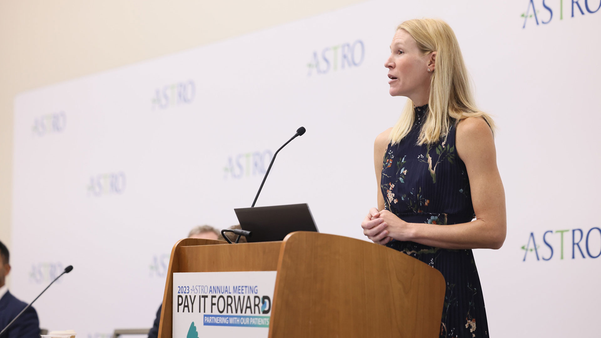
Improving palliative cancer treatment with existing diagnostic scans: Study reveals promising results
A recent study from London Health Sciences Centre and Lawson Health Research Institute suggests that using existing diagnostic CT scans in planning simple palliative radiation treatments can significantly cut down the waiting time for urgent treatment, resulting in a better experience for cancer patients.
“Reducing the time patients spend in a cancer centre has far-reaching benefits,” said lead study author Melissa O’Neil an Advanced Practice Radiation Therapist at London Health Sciences Centre’s (LHSC) London Regional Cancer Program (LRCP). “Faster treatment initiation means quicker relief from symptoms for patients. Utilizing existing scans is also cost-effective and frees up appointment slots or staff, allowing us to accommodate and assist more patients in need.”
Palliative radiation therapy is used to relieve symptoms in patients whose cancers cannot be cured. It’s often used when tumours cause pain, neurological issues or breathing problems such as blocked airways.
In the current standard practice, patients referred for palliative radiation typically require a CT simulation scan before starting their treatment. This scan creates 3D images that the patient's health care team uses to develop a customized radiation treatment plan. Unfortunately, this process often takes several hours, even with efforts to speed it up.
However, many of these patients have undergone previous diagnostic CT scans as part of their routine medical care. Previous research has shown that radiation oncology teams can create suitable palliative treatment plans for patients with bone and soft tissue metastases using these existing scans. This approach is less time-consuming than the more intensive simulation scans.
In the current study, O’Neil and her colleagues explored whether using existing CT scans to plan treatment before a patient arrives at the cancer centre could reduce their wait time while still ensuring appropriate care. They randomly assigned 33 patients who needed palliative radiation for tumours in their chest, abdomen or pelvis to either the standard treatment planning with on-site CT simulation scans or to treatment planning using diagnostic CT scans taken within the previous 28 days.
The study found that patients who didn't need the extra CT simulation scan spent much less time at the cancer centre on the day of their treatment – just under 30 minutes compared to nearly five hours for the others. Treatments were delivered successfully, and patient perception on time spent at the cancer centre was improved for those whose treatment planning used diagnostic CT scans taken without the previous 28 days.
"For patients who need radiation to help treat symptoms of cancer, it's important for us to get them treated quickly and to minimize the time they spend waiting for medical appointments,” said Dr. David Palma, Radiation Oncologist at LHSC and Associate Scientist at Lawson. “This trial shows that this new approach not only saves resources by reducing the number of scans we do, but also substantially reduces the time patients spend waiting for urgent radiation.”
"These findings are incredibly promising, especially in light of the nationwide shortage of radiation therapists," said Dr. Michael Ott, Physician Department Executive for Oncology at LHSC. “Work like this has benefits that can reach far beyond London, offering more relief for patients across the country."
The findings were presented at the American Society for Radiation Oncology’s Annual Meeting on Oct. 3, 2023. This meeting is recognized globally as the leading radiation oncology scientific event, drawing more than 8,500 attendees each year.
While the study shows promise, the research team said it's important to note that using prior diagnostic scans may not be suitable for every type of cancer or patient. It depends on the specific area being treated and the technique used.
For more information, please contact:
Jessica Rabaey
Communications Consultant
London Health Sciences Centre
T: 519-685-8500 ext. 77728
Jessica.rabaey@lhsc.on.ca
About Lawson Health Research Institute
Lawson Health Research Institute is one of Canada’s top hospital-based research institutes, tackling the most pressing challenges in health care. As the research institute of London Health Sciences Centre and St. Joseph’s Health Care London, our innovation happens where care is delivered. Lawson research teams are at the leading-edge of science with the goal of improving health and the delivery of care for patients. Working in partnership with Western University, our researchers are encouraged to pursue their curiosity, collaborate often and share their discoveries widely. Research conducted through Lawson makes a difference in the lives of patients, families and communities around the world. To learn more, visit www.lawsonresearch.ca.