Ivey Eye Institute - Services
Orthoptic Services
Orthoptics involves the correction of visual disturbance or impairment, such as those associated with strabismus or crossed eye, using various types of special lenses as well as exercises.
An orthoptist is a person who specializes in the study of the eye muscles. The orthoptist participates in the diagnosis and nonsurgical treatment of patients with decreased vision and misaligned eyes.
There are three clinical orthoptists within the department, who also conduct their own research.
Diagnostic Services
The department has a fully staffed diagnostic unit, with a full range of equipment, including fundus, slit lamp and external photography, electroretinography, electro-oculography, fluorescein angiography, specular microscopy, orbscan, optical coherence tomography, pachymeter, visual field studies, ultrasonography (both A- and B-mode) of the eye and orbit, IOL Master Lenstar.
For more information, please call St. Joseph's Hospital
Phone: 519 646-6100 ext. 66018.
Automated visual field testing
Visual field testing is a test which maps out a patients area of sight. The purpose is to determine the dimmest light visible in the patient’s peripheral vision. The patient is seated at a machine with one eye patched. A series of lights which vary in intensity are “flashed” on a screen in front of the patient. The patient then responds when they see a light by pushing a button. By compiling these responses a “map” of the patients vision is then produced. This testing is most commonly used for glaucoma patients. It is also used to document other visual defect causing conditions.
Intravenous fluorescein angiography (IVFA)
IVFA is a diagnostic test which is used to document and diagnose various diseases of the parts of the eye called the retina and choroid. The patient is seated at a special camera, and a dye called Sodium Fluorescein is injected into a vein either in the arm or hand. When the dye reaches the eyes the technician operating the camera will start taking a series of photographs. There is a bright flash of light each time a picture is taken. The photographs are often used for treatment purposes giving the physician a very accurate picture of the status of the blood vessels in the back of the eye.
Potential acuity meter (PAM)
The potential acuity meter test is used to see how well the most sensitive part of the retina called the macula is functioning. The PAM machine shines an eye chart onto the retina, and the patient is asked to read as much of the eye chart as possible. This test is often used as an indicator of how well a patient would see after cataract surgery if that surgery was indicated.
Diagnostic B-scan
A diagnostic B-scan is an ultrasound examination, which provides a two-dimensional image of the eye and surrounding orbit. High frequency sound waves are transmitted and received through a probe, which can either be placed directly on the eye or the eyelid. As the sound waves pass through the different eye tissues, echoes or reflected signals are converted into electrical energy. These signals are displayed as “bright spots” on an oscilloscope or screen.
A B-scan is performed to image parts of the eye that are not visible to the ophthalmologist. A common phrase sometimes used by the ophthalmologist is, “it is like seeing with sound.”
Conditions such as a vitreous hemorrhage, dense cataract, or scarred cornea make it impossible for the ophthalmologist to examine the back part of the eye, where the vitreous, the retina and the eye wall are located. In some cases of eye injury or trauma, where the eye is penetrated by an object, a B-scan would be performed to rule out the presence of a foreign body and also to see the extent of the damage in the eye.
A B-scan, along with a diagnostic A-scan, is used to document and differentiate eye lesions or tumours. Accurate measurements can be made to determine the size and height of an ocular tumour, as well as the shape and contour. It is also possible to make out the different types of tissue, which can help in the diagnosis of a benign or a cancerous tumour, and even if it is a cancerous tumour that has spread from its original site.
An ocular ultrasound usually takes about 10-15 minutes to perform. The patient is in a semi-reclining position in an examining chair, and most B-scans are performed by placing the probe directly on the eyelid. A small amount of coupling gel is used on the eyelid to ensure good contact between the ultrasound probe and the patient’s eyelid. The patient is asked to move their eye in different directions throughout the scan so the entire eye area can be thoroughly examined.
There is virtually no discomfort experienced by the patient unless the eye has undergone recent eye surgery or has been injured.
Orbscan iTace
The Orbscan iTrace utilizes optical technology and computer software to measure and analyze the anterior corneal curvature and elevation, posterior elevation and corneal thickness. From this information, a topographical colour map of the cornea can be created. The data is helpful to evaluate and plan to correct astigmatism, monitor corneal disease and detect irregularities in the shape of the cornea. Accurate measurement of astigmatism is important for refractive surgery, contact lens fitting and calculating the power of intraocular lenses. It is also used often to diagnose keratoconus and other corneal diseases, and corneal changes after keratotomy and keratoplasty.
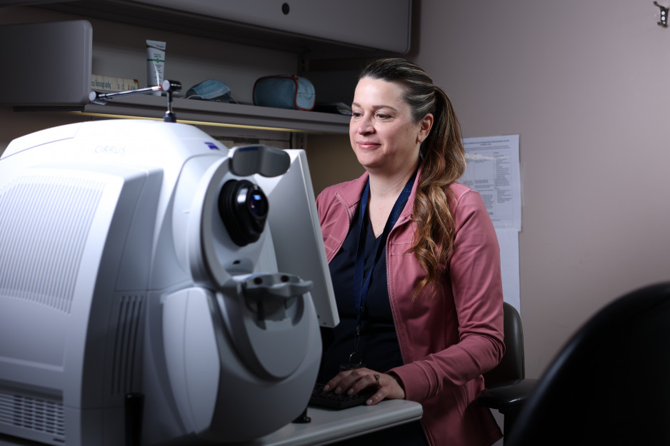
Optical coherence tomography (OCT)
Optical coherence tomography (OCT) is a noninvasive, noncontact, imaging technology which can visualize retinal structures with a resolution of 10 to 17 microns. Non-invasive OCT examinations produce real-time cross-sectional images of retinal tissue, in less than 10 minutes. OCT's high resolution, which is 10 times greater than magnetic resonance imaging (MRI) or ultrasound, may enable the detection of early microscopic signs of disruption in tissues for treatment. The ophthalmic applications may benefit patients with glaucoma as well as retinal and macular diseases. It may also be useful in some cases of those individuals considering corneal and refractive surgery.
Pachymeter
The pachymeter is an instrument that measures the thickness of the cornea and anterior chamber. Measurements can be done using ultrasonic or optical technology. The test is useful in monitoring the progression of certain disorders that cause the cornea to become thickened resulting in a loss of vision. Pachymetry is also performed to determine whether the cornea is strong enough for certain surgical procedures. It is also useful in certain patients who have glaucoma or who are glaucoma suspects.
Electrophysiology
Electrophysiology is the electrical phenomenon associated with a physiological process such as vision, brain activity or heart conduction. Specific tests can be used to evaluate visual functions.
Visual evoked potential / response (VER)
VER is a computerized recording of electrical activity at the back of the brain (occipital cortex) that results from stimulation of the retina. This potential can be elicited by several methods, including a reversing checkerboard pattern, a stroboscopic flash or a flash generated by matrix of light emitting diodes (LED).
The VEP consists of a large biphasic waveform, which occurs at 250 msec. after the onset of a visual stimulus. There are three electrodes attached to the patient’s head -one on the forehead, one at the back of the head and one on the centre of the scalp. Several readings are taken and computer averaged.
Visual Evoked Potentials assesses the central retina, optic nerve and visual pathways, and may reveal the presence of gross retinal dysfunction, demyelinating disease, e.g. multiple sclerosis (M.S.) or compressive lesions (tumors) affecting the optic nerve.
Electroretinogram (ERG)
Electroretiography (ERG) assesses the health of the retina and allows individual analysis of the special cells that allow us to see colour and light. There are two major types of cells in the retina, called cones, which are light-sensitive retinal receptor cells that provide sharp visual acuity and color discrimination, and rods (light-sensitive retinal receptor cells, which are specialized to work at low light levels or night vision).
To carry out this test the pupil of the eye must be dilated by using special eye drops. Skin-type electrodes are placed on the patient’s forehead and earlobe, and then the patient sits in a completely dark room for 30 minutes wearing special eye patches. This is called “dark-adapting” and is necessary to ensure maximum response of the rod function. At the end of the 30-min dark adaptation, topical anesthetic drops are placed in the eye and a special fibre electrode is placed inside the patient’s lower eyelid. The test entails the patient watching several different colours of light being flashed, which stimulates the rods and cones in the retina.
This test takes approximately 1-½ hours in total.
Ocular ultrasound - A-scan, axial eye length measurement
Axial eye length A-scan or biometry is the most common type of ocular ultrasound performed today. An axial eye length reading (measurement taken along the visual axis between the cornea and retina) and keratometry readings (measurement of the corneal curvature) are necessary for intraocular lens calculations. These calculations help determine the correct power or strength of lens necessary for the eye to restore vision to near normal after cataract surgery. Some patient’s also require additional glasses, mostly reading glasses.
An intraocular lens is an artificial lens implanted in the eye during cataract surgery in the same location as previously occupied by the natural lens.
These measurements take approximately 15 - 20 minutes and are usually preformed during the “pre-op” visit or assessment prior to the cataract surgery.
The technician will instill a drop of topical anesthetic in the eye(s) to numb the corneal surface and then position the patient’s head comfortably. Some readings are performed where a patient is positioned in a slit lamp type of apparatus and some measurements are done with the technician holding the A-scan probe in his or her hand. *The probe is then brought close to the patient’s eye and it gently touches the cornea where a reading or axial eye length measurement is obtained. Several readings are necessary as these measurements need to be extremely accurate.
*The patient is asked to fixate on a red target light found at the end of the A-scan probe.
Ocular coherence tomography (OCT)
Glaucoma is a progressive disease of the optic nerve that can lead to permanent loss of vision. Clinical studies have confirmed that early detection and treatment of glaucoma can slow or prevent the progression of permanent optic nerve.
There is state of the art technology that measures and analyzes the optic nerve in a more precise way and allows for detection of subtle changes over time. One of these technologies is called ocular coherence tomography (OCT). This test takes digital images of the back of your eye and computers evaluate the images to provide information about your glaucoma.
Data from this test may help to determine when treatment is necessary, and if you are already on treatment, whether it is adequate. This test will most likely need to be repeated on a regular basis. This test is completely safe, causes you no discomfort, and take only minutes to complete.
At present OHIP does cover OCT.
It is your choice to have this test. It is recommended, but should you decide to decline this test, the standard of care will continue to be delivered to you and every effort will be made to manage your glaucoma in the best manner possible.
Cataract Surgery
 St. Joseph’s Ivey Eye Institute is a world renowned leader in the provision of eye care including cataract surgery. Patients who are considering or have been booked for cataract surgery will find information below including:
A guide to cataract surgery at St. Joseph's Hospital can be downloaded from the Patient Resources section, which you will receive before your surgery in hard copy.
Find referral information for how to access cataract surgery.
Low Vision Clinic
Make the most of your existing sight: low vision services
Vision loss that cannot be corrected by ordinary glasses, contact lenses, medication or surgery is called "Low Vision". Low vision services do not restore lost vision but rather utilize the remaining vision to its fullest potential. Low Vision Services include a functional eye examination and an evaluation of how your remaining vision functions in day-to-day living.
The low vision exam evaluates not only how well you see an eye chart, but also how well you see faces, street signs, newspaper print, and all the other visual clues that guide you through the day. Afterward, low vision devices will be prescribed based on your specific needs and interests so you can continue to read and function as well as possible, and continue to enjoy hobbies and recreational activities. As part of our services, we will also teach you how to use these devices correctly so you can successfully integrate them into your daily routine.
Signs of low vision:
- Difficulty recognizing a familiar face
- Difficulty reading: print appears broken or distorted
- Difficulty seeing objects and potential obstacles such as steps, curbs, walls and furniture
- Loss of visual acuity or sharpness
- Loss of peripheral/side vision
- Extreme difficulty with light or glare
Common causes of low vision:
- Macular degeneration
- Glaucoma
- Diabetic retinopathy
- Stroke
- Corneal degeneration
- Retinitis pigmentosa
- Brain tumors, brain injuries
- Optic atrophy
- Albinism
- Inoperable cataract
Assistive Devices Program
Depending on the visual function of your eyes, the cost of low vision devices prescribed at the Low Vision Clinic may be partially funded by the Assistive Devices Program (ADP) of the Ontario Ministry of Health and Long-Term Care. However, the low vision examination fees are not covered by the Ontario Health Insurance Plan (OHIP).
Patient Resource and Education Room
The Marjorie Jean Jolliffe Resource Centre is the Ivey Eye Institute Registration Area. It contains patient resources and education on eye conditions, diseases, and disorders.
This resource space is funded by the Marjorie Jean Jolliffe Fund. Marjorie Jolliffe named the Ivey Eye Institute in her will in appreciation for the care she received from Dr. Larry Allen and the team of care givers at the Institute.
Please feel welcome to take any information material from the the Marjorie Jean Jolliffe space.