Medical Imaging: CT and CAT
Computed tomography/computerized axial tomography
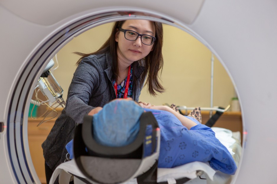
A computed tomography (CT) or computerized axial tomography (CAT) scan is a special type of x-ray that can produce detailed pictures of structures inside your body. A CT scanner works by directing a series of x-rays through your body to produce a detailed picture or a "slice" of the area being studied.
Each x-ray pulse lasts only a fraction of a second, and it takes only a few seconds for the machine to record many images.
Why is it done?
A CT scan is used to obtain information about many parts of the body, including:
- Most organs (liver, pancreas, intestines, kidneys, adrenal glands, lungs, or heart)
- Blood vessels
- Abdominal cavity
- Bones (particularly the spine) and the spinal cord
Cardiac CT angiogram, CTA or cardiac CT, uses advanced CT technology with a contrast dye (intravenous contrast material that contains iodine) to obtain detailed images of your heart muscle, coronary arteries, pulmonary veins, the thoracic aorta and the sac around your heart (pericardium).
Detailed images of the heart
During cardiac CT, 2D and 3D images are produced that allow your physician to determine whether plaque or calcium deposits are present in the artery walls.
Cardiac CT is a non-surgical method for detecting blockages in the coronary arteries. It can be done more quickly than cardiac catheterization, with potentially less risk and discomfort, and minimal recovery time.
